


Seizure disorder and thyroiditis
Students much review the case study and answer all questions with a scholarly response using APA and include 2 scholarly references. Answer both case studies on the same document and upload 1 document to Moodle.
Case Study 3 & 4 Seizure Disorders & Thyroiditis
Case Studies will be uploaded to Moodle and put through TURN-It-In (anti-Plagiarism program)
Turn it in Score must be less than 50% or will not be accepted for credit, must be your own work and in your own words. You can resubmit, Final submission will be accepted if less than 50%. Copy paste from websites or textbooks will not be accepted or tolerated. Please see College Handbook with reference to Academic Misconduct Statement.
Seizure Disorder Case Study
A 12-year-old boy began to complain of frequent headaches 4 months before his hospital admission. On the day of his admission, he had a major motor seizure, which his parents observed. During the seizure he lost bladder and bowel control. On physical examination he appeared to be in deep postictal sleep. He had no focal neurologic signs. On examination of the optic fundi, no evidence of papilledema was found. Studies Results Routine laboratory work Within normal limits (WNL) Skull X-ray study, p. 1062 No evidence of skull fracture Lumbar puncture, p. 651 Opening pressure 250 cm H2O (normal: <200 cm H2O) Closing pressure 220 cm H2O (normal: <200 cm H2O) Cerebrospinal fluid (CSF) examination, p. 651 Blood Negative Color Clear Cells Lymphocytes 0-2/mm3 (normal: <5/mm3 ) Polymorphonuclear leukocytes None (normal: none) Protein 120 mg/dL (normal: 15-45 mg/dL) Glucose 50 mg/dL (normal: 50-75 mg/dL) Cytology Questionably malignant cells Serologic test for venereal disease Negative (normal: negative) Electroencephalography (EEG), p. 549 Focal slowing of wave pattern in posterior aspect of the cerebrum (normal: regular, rhythmic, electrical waves) Brain scan, p. 785 Increase in radioactivity in the posterior aspect of the brain (normal: homogenous and minimal uptake of radioactive material) Cerebral angiography, p. 988 Neovascularity (tumor vessels) in the posterior aspect of the brain, involving the cerebellum and the occipital lobe of the cerebrum (normal: normal carotid vessels and terminal branches) Magnetic resonance imaging (MRI) of the brain, p. 1106 Tumor of the cerebellum extending into the posterior cerebrum Computed tomography (CT) scan of the brain, p. 1026 A soft tissue mass arising out of the cerebellum and invading the occipital lobe of the cerebrum Case Studies 2 Diagnostic Analysis The skull X-ray study ruled out the possibility of a skull fracture as the cause of the boy’s problem. Lumbar puncture excluded the possibility of meningitis or subarachnoid hemorrhage; however, the high protein count and questionable positive cytology indicated a possible neoplasm. An EEG located an area of nonspecific abnormality in the posterior aspect of the brain. Brain scanning, cerebral angiography, and CT scanning indicated a posterior fossa tumor. These tests are mentioned in this case study mostly for historical interest. Under most circumstances, this young boy would have a MRI of the brain early in the diagnostic period. Because of these findings, the patient underwent a craniotomy. In many centers, this young boy would have a nonoperative stereotactic brain biopsy instead of a craniotomy. An invasive medulloblastoma was found to be arising from the patient’s cerebellum and involving the occipital lobe of the cerebrum. The tumor was unresectable. Postoperatively, the patient was given phenytoin (Dilantin) and radiation therapy to the involved area. A chemotherapy regimen was administered. The patient’s tumor did not respond to the therapy, and he died 4 months after the onset of disease. Critical Thinking Questions 1. What are the major assessments that the nurse should make during seizure activity? 2. Why is the EEG a priority study for patients with seizure disorders?
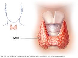
Thyroiditis Case Study
The patient, a 23-year-old woman, has had a bout of flulike symptoms over the past few weeks. Most recently, she has become increasingly tired. She is taking birth control pills to control her menses. Her anterior neck became painful during the past few weeks. The physical examination results reveal that her thyroid is diffusely enlarged and mildly tender. Studies Results Routine laboratory tests Within normal limits (WNL) Total thyroxine (T4), p. 497 8 mcg/dL (normal: 5-12 mcg /dL) Free T4 0.5 ng/dL (normal: 0.8-2.7 ng/dL) Free T4 index 0.4 ng/dL (normal: 0.8-2.4 ng/dL) Triiodothyronine (T3), p. 506 52 ng/dL (normal: 70-205 ng/dL) Thyroxine-binding globulin (TBG), p. 495 12 mg/dL (normal: 1.7-3.6 mg/dL) Thyroid stimulating hormone (TSH), p. 486 32 microunits/mL (normal: 2-10 microunits/mL) Thyroid scanning, p. 839 Enlarged gland; normal shape, position, and function of the thyroid gland. No areas of decreased or increased uptake Thyroid ultrasound, p. 895 Enlarged gland; normal shape and position of the thyroid gland Thyroid antibodies Antithyroglobulin antibody, p. 102 1:250 (normal: titer <1:100) Antithyroid peroxidase antibody, p. 104 1:500 (normal: titer <1:100) Thyroid-stimulating immunoglobulins, p. 491 Negative Diagnostic Analysis Total T4 measures protein-bound and unbound T4. Because the patient was taking birth control pills, her TBG was elevated; therefore, her total T4 was normal. Free T4 and FT4 index tests measure unbound T4. When the free T4 and the FT4 index were measured, they were found to be low, indicating that the patient had hypothyroidism. The TSH level was elevated because of primary failure of the thyroid. The thyroid antibodies were elevated, indicating that the patient had Hashimoto thyroiditis. Her long-acting thyroid stimulator (LATS) levels were normal, discounting Graves disease as a cause of her diffusely enlarged thyroid. Her thyroid ultrasound and scan failed to show any localized, defined tumor. The patient was started on thyroid replacement therapy, and her TSH level returned to normal. Over the next few weeks, she felt markedly better. Her thyroid pain and tiredness disappeared. Critical Thinking Questions 1. Why were the thyroid antibodies important in this patient’s diagnosis? 2. What symptoms might she experience if too much thyroid replacement medication were administered?
We can write this or a similar paper for you! Simply fill the order form!