Pathophysiological Presentation of DVT and CVI Order Instructions: Advanced practice nurses often treat patients with vein and artery disorders such as chronic venous insufficiency (CVI) and deep venous thrombosis (DVT).

While the symptoms of both disorders are noticeable, these symptoms are sometimes mistaken for signs of other conditions, making the disorders difficult to diagnose. Nurses must examine all symptoms and rule out other potential disorders before diagnosing and prescribing treatment for patients. In this Assignment, you explore the epidemiology, pathophysiology, and clinical presentation of CVI and DVT.
To prepare:
Review the section “Diseases of the Veins” (pp. 585–587) in Chapter 23 of the Huether and McCance text. Identify the pathophysiology of chronic venous insufficiency and deep venous thrombosis. Consider the similarities and differences between these disorders.
Select a patient factor different from the one you selected in this week’s Discussion: genetics, gender, ethnicity, age, or behavior. Think about how the factor you selected might impact the pathophysiology of CVI and DVT. Reflect on how you would diagnose and prescribe treatment of these disorders for a patient based on the factor you selected.
Review the “Mind Maps—Dementia, Endocarditis, and Gastro-oesophageal Reflux Disease (GERD)” media in the Week 2 Learning Resources. Use the examples in the media as a guide to constructing two mind maps—one for chronic venous insufficiency and one for venous thrombosis. Consider the epidemiology and clinical presentation of both chronic venous insufficiency and deep venous thrombosis.
To complete:
Write a 2- to 3-page paper that addresses the following:
Compare the pathophysiology of chronic venous insufficiency and deep venous thrombosis. Describe how venous thrombosis is different from arterial thrombosis.
Explain how the patient factor you selected might impact the pathophysiology of CVI and DVT. Describe how you would diagnose and prescribe treatment of these disorders for a patient based on the factor you selected.
Construct two mind maps—one for chronic venous insufficiency and one for deep venous thrombosis. Include the epidemiology, pathophysiology, and clinical presentation, as well as the diagnosis and treatment you explained in your paper.
This Assignment is due by Day 7.
Note: The School of Nursing requires that all papers submitted include a title page, introduction, summary, and references. The Sample Paper provided at the Walden Writing Center provides an example of those required elements (available at http://writingcenter.waldenu.edu/57.htm). All papers submitted must use this formatting.
VERY IMPORTANT SEE BELOW FOR THE GUIDE FOR THE MIND MAP
Media
Zimbron, J. (2008). Mind maps—Dementia, endocarditis, and gastro-oesophageal reflux disease (GERD) [PDF]. Retrieved from http://www.medmaps.co.uk/beta/
Gastro-oesophageal reflux disease. [Image]. Used with permission of MedMaps.
This media provides examples of mind maps for dementia, endocarditis, and gastro-oesophageal reflux disease (GERD).
Pathophysiological Presentation of DVT and CVI Sample Answer
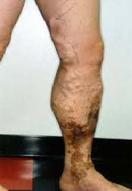
Chronic Venous Insufficiency (CVI) arises due to the incompetence of vascular walls as well as valves of the veins. This disorder leads to a reduction in blood flow to the heart resulting in pooling of blood or stasis in the extremities especially the lower limbs. Patients with CVI usually complain of pain and swelling in the limbs. Conversely, deep venous thrombosis (DVT) arises when clotting occurs in the deep veins in the lower limbs (Patel & Brenner, 2013). Patients suffering from DVT usually complain of pain as welling as swelling just as those with CVI. The presentation of these conditions is almost similar. It is for this reason that health care providers take extra caution when diagnosis CVI and DVT.
The Pathophysiological Presentation of DVT and CVI
The key pathophysiological difference between CVI and DVT is that DVT occurs in deep veins whereas CVI occurs majorly in superficial veins. CVI affects popliteal, femoral, and peroneal veins while DVT mail affects the soleal vein. Chronic Venous Insufficiency arises as a result of damage of the endothelial walls and valves in the veins (Eberhardt & Raffetto, 2014). Some of the common causes of CVI include pelvic tumors, DVI, and vascular malformations. The valves of patients suffering from CVI are incompetent in that they cannot hold blood back against the force of gravity. Consequently, blood pools in the lower extremities leading to swelling especially in the ankles and the legs. Moreover, individuals with CVI present with venous stasis ulcers, varicose veins, pain the feet, and itching and flaking of the skin. On the other hand, DVT develops due to clotting in the veins. Severe clinical complications occur when the formed clots lyse and get into the general circulation. Blood from deep veins usually flows into the lungs. Therefore, when this blood carries clots with it, it may lodge them in the lungs causing pulmonary embolism, one of the most severe result of DVT (Goldhaber & Bounameaux, 2012). Often CVI presents with dermatitis and ulceration due to the structural difference between the deep veins and superficial veins. That is, the superficial veins have an adipose layer and a connective tissue whereas the deep veins have a fascia and muscles. This gives deep veins more protection and structural support.
Venous and arterial thrombosis have a number of similarities although they differ in terms of their pathophysiology, clinical interventions, and epidemiology. Venous thrombosis occurs in undamaged parts of venous walls and in areas that have low sheer pressure. This disorder leads to the formation of red thrombi. Conversely, arterial thrombosis occurs in parts that have high sheer stress and are rich in plaques. Unlike, venous thrombosis, arterial thrombosis forms white thrombi.
Patient Behavior
The predisposition and pathophysiological advancement of DVT and CVI relies heavily on the lifestyle of an individual. The pathophysiology of DVT and CVI is enhanced when a person engages in activities that enhance the metabolic syndrome. Some of the most notable practices that have been cited to predispose individuals to CVI and DVT include lack of physical exercises, smoking, intake of meals rich in cholesterol, and psychosocial behavior (Csordas & Bernhard, 2013). Smoking affects the circulation of blood and enhances blood clotting. On the other hand, inactivity such as sitting for long periods causes calf muscles to contract hence inhibiting the proper circulation of blood. Lack of activity may also result in an increase of weight which then increases pressure in veins especially in the legs and the pelvis.
When diagnosing of CVI and DVT based on behavior, a physician should enquire the social history of the patient. For instance, s/he can ask the patient whether s/he smokes or has ever smoked. If the patient smokes, he should enquire when the patient started smoking and how many sticks he smokes in a day. Questions on whether the patient engages in physical exercises such as jogging or long distance travelling are also essential in finding a differential diagnosis.
Clinical interventions for these patients involves the use of pharmacological as well as non-pharmacological approaches. If the patient smokes, a physician should assess the willingness of the patient to quit smoking. If s/he is willing to make a quit attempt, a brief counselling session should be introduced, medications such as bupropion will be offered as well as self-help resources. Follow-up visits should also be scheduled. The patient should also be advised to engage in physical exercises such as jogging. The patient should also limit his/her intake of cholesterol, a leading factor in DVT development.
Mind Maps
|
||||||||||||||
|
||||||||||||||
|
||||||||||||||
|
||||||||||||||
|
||||||||||||||
|
||||||||||||||
Pathophysiological Presentation of DVT and CVI References
Berkman, L. F., Kawachi, I., & Glymour, M. M. (Eds.). (2014). Social epidemiology. Oxford University Press.
Csordas, A., & Bernhard, D. (2013). The biology behind the atherothrombotic effects of cigarette smoke. Nature Reviews Cardiology, 10(4), 219-230.
Eberhardt, R. T., & Raffetto, J. D. (2014). Chronic venous insufficiency.Circulation, 130(4), 333-346.
Goldhaber, S. Z., & Bounameaux, H. (2012). Pulmonary embolism and deep vein thrombosis. The Lancet, 379(9828), 1835-1846.
Patel, K., & Brenner, B. (2013). Deep venous thrombosis. Medscape reference.


