THE USE OF ESTROGENS and PROGESTINS and the RISK of BREAST CANCER in POSTMENOPAUSAL WOMEN
Order Instructions:
Write a 2- to 3-page word document to complete the following assignment tasks.
•Choose the research article you selected in W1 Assignment 3. Describe the sample, demographics, data collection process, setting, and the instrument, tool, or survey used in the article.
•Discuss the author’s summary about the validity and reliability of the instrument.
Cite all sources in APA format.
SAMPLE ANSWER
THE USE OF ESTROGENS and PROGESTINS and the RISK of BREAST CANCER in POSTMENOPAUSAL WOMEN
Description of the sample:
This prospective cohort study is an extension of a 1976 Nurses’ Health Study where patients 30-55 years of age were recruited and followup every 2 years from 1978 for 12 years. From this cohort, a non-randomized sample of 50-69 years old postmenopausal women (all state registered nurses) were followed for additional two years through to 1992. Participants were selected for followup based on the inclusion / exclusion criteria established from the 1976 questionnaires and baseline information on risk factors for breast cancer, including hormone therapy and subsequent menopausal status. Out of 725,550 women who were followed up, 1935 of them developed invasive breast cancer. Premenopausal women, as well as those who reported a breast cancer incident or other cancer apart from nonmelanoma skin cancer at recruitment were excluded from subsequent followup.
The process of data collection:
Data was collected through the use of questionnaires and telephone interviews, commencing from 1976 when the study was initiated and through to 1992. The group of interest was postmenopausal women, who had been diagnosed with breast cancer and were currently undergoing hormone therapy. In order to determine any associations between the occurrence of breast cancer and hormones, data were stratified and further analysed by a multivariate analysis as a means of controlling the possible confounding effects that might be caused as a result of other risk factors.
The demographics of the study population was as follows:
Origin/Sex/Education: All participants were US-based female state registered nurses.
Age: Study participants consisted mainly of postmenopausal women between the ages of 50 and 69 years, who were followed up every second year from 1976 to 1992.
Menopause status: This included the age of menopause onset and the type of menopause; whether women had a natural onset of menopause, had had hysterectomy but no bilateral oophorectomy or had had hysterectomy with bilateral oophorectomy, as well as the smoking status. In addition, women were furthered classified based on a breast cancer diagnosis, a history of cancer in the family, a family history of benign breast cancer and the time period.
Hormone use status: The hormone status included the duration of hormone use, the preparation of hormones used (use of progestin alone, conjugated estrogen alone, estrogen and progestin of estrogen and testosterone) and the dose. The cut off for the duration of hormone use related to high cancer risk was at 5 years.
Setting: The study was performed in the United States.
Instrument, tool, or survey used in the study.
Age standardized mammographic screenings were performed for breast cancer diagnosis.
Interviews at followup were done by telephone and by the use of structured questionnaires mailed to participants.
When participants did not return questionnaires, the National Death Index was used to ascertain the death of the nonrespondent cases.
Hospital records and pathology reports were used to confirm data on cancer diagnoses.
Validity and reliability of the instrument
Even though self-reporting and sampling questionnaires done by mail could be subjectively biased (Phellas, Bloch & Seale, 2012), the authors were confident in the accuracy of the participants’ reports; which according to them was extremely high, as they achieved nearly 100% followup completion through questionnaires and telephone for both nonfatal and fatal breast cancer respectively. Nevertheless, the administering of questionnaires as a sampling method is generally considered cheap, having no interviewer bias and with an added benefit of anonymity (Phellas, et al., 2012). While interviews have the advantage of being direct and have a better response rate (Phellas et al., 2012), its validity and reliability is still questionable, as it is not always possible to cross check information from self-reports. In this study, 10 cases of self-reported breast cancer could not be confirmed from medical records, further stressing the subjectivity of this method. Here, the authors validated the data for breast cancer occurrence using hospital records when available and the death of participants by use of the National death Index. In any case, this method is still unreliable in the event that the records are incomplete or in situations where medical records are unavailable, thus, resulting in “missing” data, as was the case in this study; wherein records could not be obtained for 7 percent of the self-reported cases of breast cancer.
Since age-standardised mammography could detect a larger proportion of in situ breast cancers, the validity / reliability of mammograms could be questionable as a tool for acquiring data relevant to the study. However, the authors accounted for this discrepancy by excluding such cases from the data analysis. Overall, data obtained with the above tools proved relevant to the objective of the study which set out to investigate the risk of developing breast cancer in postmenopausal women on hormone therapy; specifically estrogen and progestin. A review of literature was used to corroborate facts, contradict findings or to indicate existing gaps in the literature on the subject. Meanwhile, the use of multivariate analyses further controlled for confounding factors.
Reference
Phellas, C.N., Bloch, A., & Seale, C. (2012). Structured methods: interviews, questionnaires and observation. In C. Seal (Ed.), Researching Society and Culture. Retrieved from www.sagepub.com
We can write this or a similar paper for you! Simply fill the order form!
Combine all elements completed in previous weeks (Topics 1-4) into one cohesive evidence-based proposal and share the proposal with a leader in your organization. (Appropriate individuals include unit managers, department directors, clinical supervisors, charge nurses, and clinical educators.)
Obtain feedback from the leader you have selected and request verification using the Capstone Review Form. Submit the signed Capstone Review Form to CONHCPfield@gcu.edu
For information on how to complete the assignment, refer to “Writing Guidelines” and the “Exemplar of Evidence-Based Practice Capstone Paper.”
Include a title page, abstract, problem statement, conclusion, reference section, and appendices (if tables, graphs, surveys, diagrams, etc. are created from tools required in Topic 4).
Prepare this assignment according to the APA guidelines found in the APA Style Guide, located in the Student Success Center.
This assignment uses a rubric. Please review the rubric prior to beginning the assignment to become familiar with the expectations for successful completion.
You are required to submit this assignment to Turnitin. Please refer to the directions in the Student Success Center.
Note: All Capstone Projects are to be submitted to the College. Please submit an electronic copy to this e-mail address: CapstoneRNBSN@gcu.edu
7 NRS 441v.10R. Writing guidelines.docx 8 NRS 441v.10R.Exemplar of Evidenced-Based Practice.docx NRS441V.R.CapstoneReviewForm_1-27-14.docx
SAMPLE ANSWER
Abstract
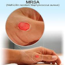
The frequency of people with MRSA infections has increased considerably in recent years. In 2006, over 50% of all cases of skin infections because of MRSA happened in healthy persons living in the community. The 3 types of MRSA include healthcare-associated MRSA, hospital-associated MRSA, and community-associated MRSA. In the year 2008, MRSA resulted in about 89,786 cases of invasive disease leading to nearly 15,300 deaths in America. In the year 2008, roughly 27 percent of hospital-acquired MRSA infections were because of USA300 strains. MRSA is a major threat to communities and to patients in healthcare facilities. An MRSA infection can actually be more severe compared to other bacterial infections and can be life threatening. In America, studies indicate that MRSA is actually responsible for about 60 percent of community acquired infections with S Aureus presenting to healthcare facilities. The rates of MRSA is increasing rapidly in many regions and there is a dynamic spread of strains all over the world. At present, healthcare associated/acquired MRSA (HA-MRSA) is endemic in hospitals. The proposed solution for the prevention of MRSA is to provide education to individuals and communities on the ways to prevent the spread as well as transmission of the difficult-to-treat MRSA. The main reason for providing education to communities and individuals is essentially to promote health and prevent disease.
Problem Statement
MRSA is defined as an oxacillin minimal inhibitory concentration of at least 4 µg/mL (Raygada & Levine, 2010). The rates of MRSA keep on increasing in many countries around the world. Romano, Lu and Holtom (2011) stated that MRSA infections occur in 3 particular groupings of people: (i) those with recent hospitalization or continuing contact with dialysis units, medical clinics, or those who are going through intricate outpatient treatments, for instance chemotherapy. They are exposed to healthcare-associated MRSA. (ii) Those who are presently within the hospital setting, and these are exposed to hospital-associated MRSA. (iii) Those in the community and these are exposed to community-associated MRSA (Green et al., 2012). A person can become colonized, meaning to be infected with MRSA, by touching a surface which is contaminated, for instance a phone, a door handle, or a counter top; and by touching the skin of an individual colonized with MRSA (Raygada & Levine, 2009).
Mascitti et al. (2010) stated that Staphylococcus is a significant public health issue, and is known to be associated with infections that are difficult to treat. It is also linked to high incidences of mortality and morbidity, as well as increased costs of health care. Staphylococcus is essentially a bacterium which is carried on the nasal lining or skin of about 30% of healthy people (Stefani et al., 2012). In such settings, the bacteria usually does not cause any symptoms, and in such instances the individual is colonized with MRSA. Nonetheless, when the skin of that person is damaged, for instance is cut or scratched, this bacterium can bring about various problems ranging from severe illness to a mild pimple, particularly in elderly persons, children, and persons whose immune system is weakened (Koydemir et al., 2011). Methicillin-resistant staphylococcus aureus is a serious threat to the community and to patients in healthcare facilities. It is particularly difficult and expensive to treat because of its resistance to common antibiotics.
In the year 2006 in America, there were roughly 94,350 invasive MRSA infections, resulting in over 17,900 deaths annually (Green et al., 2012). In America, the proportion of hospital-acquired MRSA infections is high. From 2009 to 2010, 58.7 percent of S.aureus catheter-associated urinary tract infections, 54.6 percent of S. aureus central line associated bloodstream infections, 43.7 percent of S. aureus surgical site infections, and 48.4 percent of S. aureus ventilator-associated pneumonia episodes were caused by MRSA (Calfee et al., 2014). In the year 2008, MRSA resulted in about 89,786 cases of invasive disease leading to nearly 15,300 deaths in America (Prosperi et al., 2013). In the year 2008, roughly 27 percent of hospital-acquired MRSA infections were because of USA300 strains.
Community-associated MRSA was initially seen as a cause of infection in community-based people without any health care contact. The emergence of Community Acquired-MRSA as a cause of hospital acquired infections places many patients, health workers, as well as their community contacts possibly at risk of getting an MRSA infection (Otter & French, 2011). The emergence of community-associated MRSA also serves to expose its strains to the selective pressure of antibiotic usage in hospitals possibly leading to increased anti-biotic resistance. Different strains of CA-MRSA have invaded healthcare settings. In the year 2008, roughly 27 percent of hospital-acquired MRSA infections were because of USA300 strains. Currently, MRSA strains are resistant to the available β-lactam antibiotics, such as cephalosporins and penicillins. Gray (2014) pointed out that Methicillin-Resistant Staphylococcus Aureus are commonly not just resistant to methicillin and other β-lactam antibiotics, but they are also resistant to other classes of antibiotics.
MRSA is a major threat to communities and to patients in healthcare facilities. An MRSA infection can actually be more severe compared to other bacterial infections and can be life threatening. There is a growing occurrence of health care associated infections with MRSA in youngsters with underlying conditions predisposing to infection with S aureus. In America, studies indicate that MRSA is actually responsible for about 60 percent of community acquired infections with S. Aureus presenting to healthcare facilities (Gray, 2014). According to Stefani et al. (2012), the rates of MRSA is increasing rapidly in many regions and there is a dynamic spread of strains all over the world. At present, healthcare associated/acquired MRSA (HA-MRSA) is endemic in hospitals. The proposed solution for the prevention of MRSA is to provide education to individuals and communities on the ways to prevent the spread as well as transmission of the difficult-to-treat MRSA. The main reason for providing education to communities and individuals is essentially to promote health and prevent disease. The education activities would be targeted at healthcare workers and the community members in order to prevent community-associated MRSA, healthcare-associated MRSA, and hospital-associated MRSA. One of the most important ways of protecting community members, healthcare workers, and patients is by providing education both to patients and community members.
Conclusion
Methicillin-resistant staphylococcus aureus is a serious threat to the community and to patients in healthcare facilities. It is particularly difficult and expensive to treat because of its resistance to common antibiotics. In the year 2006 in America, there were roughly 94,350 invasive MRSA infections, resulting in over 17,900 deaths annually. There is a worldwide epidemic of CA-MRSA and different strains of CA-MRSA are emerging as a cause of healthcare-associated infections and hospital outbreaks have taken place all over the world. As an emerging cause of hospital-acquired infections, CA-MRSA puts many healthcare workers and patients potentially at risk of developing MRSA infection.
References
Calfee, D. P., Salgado, C.D., Milestone, A.M., Harris, A.D., Kuhar, D.T., Moody, J…Yokoe, D.S. (2014). Strategies to prevent Methicillin-resistant staphylococcus aureus transmission and infection in acute care hospitals: 2014 Update. Infection Control and Hospital Epidemiology, 35(7), 52-9. Retrieved from http://www.jstor.org/stable/10.1086/676534
Gray, J. W. (2014). MRSA: The problem reaches pediatrics. Archives of Disease in Childhood; 89: 297-298. Retrieved from http://adc.bmj.com/content/89/4/297.full
Green, B. N., Johnson, C. D., Egan, J. T., Rosenthal, M., Griffith, E. A., & Evans, M. W. (2012). Methicillin-resistant Staphylococcus aureus: An Overview for Manual Therapists. Journal of Chiropractic Medicine, 11(1), 64-76. Retrieved from http://www.ncbi.nlm.nih.gov/pmc/articles/PMC3315869/
Koydemir, C., Kulah, H., Ozgen, C., & Hascelik, G. (2011). Methicillin-resistant staphylococcus aureus biosensors for detection of Methicillin-resistant staphylococcus aureus. Biosensors and Bioelectronics, 29(1), 1-12. Retrieved from http://www.ncbi.nlm.nih.gov/pubmed/21856144
Mascitti, K. B., Gerber, J. S., Zaoutis, T., Baron, T. D., & Lautenbach, E. (2010). Preferred treatment and prevention strategies for recurrent community-associated Methicillin-resistant staphylococcus aureus skin and soft-tissue infections: a survey of adult and pediatric providers. American Journal of Infection Control, 38(4), 324-328. Retrieved from http://www.ajicjournal.org/article/S0196-6553%2810%2900063-5/abstract
Prosperi, M., Veras, N., Azarian, T., Rathore, M., Nolan, D., Rand, K., Cook, R. L., Johnson, J., Morris, G. L., & Salemi, M. (2013). Molecular epidemiology of community-Associated Methicillin-resistant staphylococcus aureus in genomic era: A cross-sectional study. Science Reports, 3(1902), 1-7. Retrieved from www.ncbi.nlm.nih.gov/pmc/articles/PMC3664956/
Romano, R., Lu, D., & Holtom, P. (2010). Outbreak of community-acquired Methicillin -resistant staphylococcus aureus skin infections among a collegiate football team. Journal of Athletic Training, 41(2), 141-145.
The patient has many presenting symptoms and this is a challenge to identifying the disease at once. The productive cough of the patient is purelent indicating the presence of a large number of white blood cells mainly neutrophillic granulocytes. The purulent sputum of asthmatic is as a result of eosinophillic cells (Farzan N.p). The patient’s blood stained sputum (hemoptysis) may be caused by a wide range of underlying disorders ranging from trauma to heart problems to lung diseases to many other infections. Patients in industrialized countries such as china, bronchitis, bronchogenic carcinoma and bronchiectasis may lead to a productive blood-stained cough but are ruled out since chest X-rays in these diseases are shown to be normal (Jaiswal pp. 176-178.). Lung cancer is also ruled out because its diagnosis does not indicate the presence of rod-like bacteria. Pulmonary embolus is also ruled since diagnosis of sputum is found to be non-purulent. Hemoptysis and purulent sputum may also be observed in patients with HIV, where the common cause is typical pneumonia (Health Grades Editorial Staff, Chamberlain 2012).
Home. n.d)
A patient with pneumonia may also present a cough with purulent sputum (Lower Respiratory Tract Infections N.p). Scanty acid-alcohol fast bacilli are also observed in a patient with HIV but this infection is also ruled since HIV patients show a normal X-ray. The patient may be suffering from Chronic Obstructive Pulmonary disease since X-ray shows flattened diaphragm but it also ruled out because the Ziehl Neelsen’s method does not show the presence of the rod –like bacteria (Acute Exacerbations of COPD, N.p). The patient may be having tuberculosis which is the most common cause of hemoptysis and also due to the observation of scanty acid-alcohol fast bacilli in the patient’s sputum (Yoon pp. 172). Observation of heavy pus cells and red blood cells and multiple light areas which coalesce provides evidence that the patient has tuberculosis (Tuberculosis, advanced – chest x-rays N.p).
Table of the Biochemistry Results
Protein
Result
Normal Range
High (),
normal () or
low ()
C-reactive protein
13mg/l
1-3 mg/l
10-40
10
1
Creatine kinase
125IU/I
10-120IU/I
308
39-308
39
C-Creatine (hs-CRP) is an essential clinical tool used by physicians to assess patients to determine if they can benefit from a statin therapy. The rise in levels of CRP is brought about inflammation hence it serves as a marker for inflammation. Different diseases and infections have varying ranges of CRP. The CRP of 13 mg/l measured for the patient indicates an increased sensitivity presenting mild inflammation.
(The Relationship Between C-Reactive Protein and Cardiovascular Disease. (n.d.)
The patient’s CK is within the normal range. This implies that there is no injury to muscles in the body (Clinical significance of markedly elevated serum creatine kinase levels in patients with acne on isotretinoin. n.d.).
Chest X-ray Pathology Test
The patient was directed to stand in front of the X-ray machine and instructed to hold his breath at the time the X-ray is taken. The radiologist usually takes two images, that is, one is taken when the patient is standing next to the machine while the other is taken while standing sideways. In case the patient is pregnant, chest X-rays are not recommended at all. In fact, no chest X-ray should be taken of the pregnant woman when she is in first six months pregnant.
(TUBERCULOSIS: No longer down and out. n.d)
All other patients can have the chest X-rays as long as they have diseases suggestive of a chest related problem. These may include the patient experiencing a persistent cough, coughing blood or productive cough with purulent, and having difficulty in breathing. If the patient has shown advanced signs of tuberculosis, one’s doctor can order for a chest X-ray. There are no expected complications as long as it is not repeated many times within a short time or a certain number of times in the patient’s life span.
The patient’s symptoms suggest that he has Tuberculosis (cough, rod-like bacteria, fever). He, therefore, would require chest X-ray besides microbiological testing. The radiologist will take both the lateral and the posterior-anterior (PA) films with each having clear notes. The typical changes he will be looking for include air space consolidation, fibrous contraction and cavitation on the superior parts of the lobes or one or both superior parts of the lower lobes or upper lobes. The extensive infiltrate with air space consolidation in noted with a bronchogram( ). The number of cavities formed is indicated by (+). Ina addition, reticulonodular satellite fibrosis and lesions are seen surrounding the involved lung which is normally identified by a traction of the right upper hilum. However, atypical finding will be more profound if the patient has human immunodeficiency virus (HIV) (Testing for tuberculosis n.d.).The result on the X-ray depends on the level of the disease. On the X-ray result film there will be seen an abnormality on the mid and lower lung fields.
Interpretation of data and Diagnosis of the patient
Based on the symptoms which have been listed, the patient has an upper respiratory infection which indicated by the cough (Rabkin N.p, n.d). There are a number of diseases which are indicated by the hemoptysis such as bronchogenic and bronchitis. But only Tuberculosis diagnoses with the rod-like bacteria in the purulent and blood-stained sputum. The patient experiences fever which comes about by the body’s struggle to fight the foreign disease causing microorganisms. That is why his average body temperature is higher than the normal 37 .4 d degrees centigrade. According to the X-ray, the arrow points to the air space consolidation, fibrous contraction and cavitation which can be observed clearly on the superior parts of the lobes or one or both superior parts of the lower lobes or upper lobes.
The suggestion that the patient is suffering from TB is based on other examinations made which indicate that the patient is not generally well. The patient was also referred to the urologist because he was suspected to have genitourinary tuberculosis. The result from the urologist which indicated 15 leukocytes in the field, confirms the presence of foreign antigens in the genitourinary system, probably, the mycobacterium, the rod shaped bacteria which causes tuberculosis (Savage pp. 1998 N.p).
More evidence that the patient is suffering from Tuberculosis is indicated by the result obtained from staining the purulent and blood stained sputum with Ziehl Neelse’s method which also confirmed the presence of large a number of scanty acid-alcohol fast bacilli. The result indicated that each field had more than 20 pus cells within a single field. This was an indication a high density of the mycobacterium, the organism behind the genesis and progression of tuberculosis. Besides pus cells, rod-like bacteria or streptococci are observed. Although few in number, they are still the causative and infectious agents of both pulmonary and genitourinary tuberculosis.
Ziehl neelsen staining. (n.d.).
The presence of red blood cells also indicates that more oxygen is needed to for respiration to produce energy needed by leucocytes. This suggestion was based on the underlying symptoms such as the immobilization of the commercial disease associated antigens. This were observed on the plastic micro-wells’ surfaces and were specifically bound IgG antibodies from Alfred’s serum to his diseased parts. There was also a positive result when peroxidise- conjugated goat which is also indicated by anti-human IgG bound Alfred’s IgG in change. Also, resistance to Amphicilin and Isoniazid in which the test carried out shows sensitivity to the drugs.
5. Discussion
a) The Lesion (Abnormality) in the Chest X-Ray
The lesion is observed as an extensive infiltrate and air-space consolidation on the upper lobe of the right lung. A number of cavities are also observed as indicated by the positive (+) signs. Also, observed are surrounding reticunodular satellite lesions which are very easy to note. There are also fibrotic lung lesions. These are also consistent with tuberculosis at an untreated inactive state of the disease. These lesions make the patient more at risk than their counterparts with a tuberculosis infection if both were more than eight years. b) Why did the urologist request the submission of three specimens?
X-ray alone is not enough to confirm that a suspect has TB . The urologist might have requested to carry out other diagnostic tests associated with TB. The results obtained support the radiology results. The comparison of the results from both departments helps the two practitioners to confirm with certainty the actual disease the patient is suffering from. The results from the urology department such as cid-fast smear which is used for microscopic identification of the cause of the pulmonary tuberculosis. The results show the presence bacilli and pus cells.
c) Significance of Using Early Morning Specimens
The urologist requested for the urine specimens produced by the patients in the early morning since it is less contaminated by lyses red blood cells. This is because the urine collected soon after a prolonged recumbency or soon after a vigorous physical or even sexual activity should not be examined when assessing the patient’s state of microhematuria since he knows it is contaminated. Patients are advised to void the first 5 ml of the urine and then collect up to 50 ml of the remaining urine in a sterile bottle.
d) Antibiotics Used As Treatment of Pulmonary TB
After diagnosing with TB, a number of antibiotics are prescribed by a doctor as a therapy for the infections and to prevent emergence of resistant bacteria in the body. They are used in a period of 6-12 months. The combination of the antibiotics includes:
Isoniazid
Rifampin
Pyrazinamide,
Ethambutol
(Tuberculosis (TB) – Treatment. n.d.).
(The rationale for recommending fixed-dose combination tablets for treatment of tuberculosis. n.d.).
e) Route; Drug Resistance; Combination Therapy and Length Of Therapy.
Since is has pulmonary TB, he will take two antibiotics orally in a combination of rifampicin and isoniazid every day for six months. He will have an additional oral administration of two antibiotics in a combination of pyrazinamide and ethambutol every day for two months (Tuberculosis (TB) – Treatment. n.d.). f) Further Investigations to Determine the Spread of the Disease
To determine the spread of the disease within him, three tests may be conducted. Positive tuberculin test leads to the development of cell-mediated immunity which develops within 2-8 weeks from the time of infection. The mechanism behind this phenomenon is that activated T Lymphocytes combine with macrophages to form granulomas which are effective in limiting replication hence curbing the spread of bacilli. Ziehl Neelsen method may be used to determine the level of infection by the bacilli. Lastly, use biochemistry tests such as determine the level of damage to specific muscles of the body. g) The Disease Progress and Prognosis for the Patient
Tuberculosis disease begins with infection and overcoming the carrier of the mycobacterium’s immune system defences. During primary tuberculosis, the disease is dormant and the immune system is able to contain the infecting and its spread. The bacteria multiplies, it continues affect the immune system and eventually overwhelms it leading to tuberculosis (Testing for tuberculosis n.d.).
Jaiswal, A., Munjal, S., Singla, R., Jain, V. & Behera, D. 2012, “A 46-year-old man with tracheomegaly, tracheal diverticulosis, and bronchiectasis: Mounier-Kuhn syndrome”, Lung India, vol. 29, no. 2, pp. 176-178.
The decision about whether or not to screen for a condition can be quite controversial. However, even in the case of noncontroversial screening programs, such as blood pressure screening, there will always be factors that argue for and against the implementation of the screening program.
In preparation for this week’s Discussion, consider the following controversial screenings: genetic screening for breast or prostate cancer, mandatory HIV screening, and obesity screening of school-aged children. Consider the pros and cons of screening for each of these health issues.
Post by Day 4 a response to the following:
Please describe the topic you selected and give some background about factors that contribute to a decision whether or not to implement the screening program within the population at large or within a subgroup of the population.
Choose and “claim” a side to argue-either pro or con-and provide an argument, supported by scholarly evidence and properly referenced, for the side you chose.
SAMPLE ANSWER
Genetic Screening for Breast or Prostate Cancer
Factors that contribute to a decision of whether or not to implement the genetic screening for breast or prostate cancer within a population of adult patients are varied. One of the factors is the history of breast or prostate cancer. According to (Caltabiano & Ricciardell (2013), breast or prostate cancer have a history of how they are transmitted and how long it takes for the bacteria to cause symptoms in the body, what happens if treatment is given, and what happens if treatment is not dispensed. Another factor that can make the disease to be screened is because it has preclinical or asymptomatic stage, whereby, the individual is diseased but is not showing symptoms (Caltabiano & Ricciardell, 2013). Breast or prostate cancer takes many years to begin to develop and, therefore, screening is recommended. The other important factor that could lead to screening of breast or prostate cancer is because the treatment that could be provided at an early stage would result in a more favorable outcome for the individual, than if the treatments were administered after symptoms appear.
Miller, Ashar, Sisson and Johns Hopkins University (2010) attest that medical practitioners may not recommend genetic screening for breast or prostate cancer because of its respective cons. One of the cons is that normal screening results do not guarantee healthy genes in that, if a patient tests negative for the breast or prostate mutations, but the presence of mutation is not confirmed in a family member with the respective cancer, the patient is still considered high risk. Another con is that close monitoring with regular tests does not always succeed in detecting breast or prostate cancer (Miller, Ashar, Sisson & Johns Hopkins University, 2010). Some patients end-up being diagnosed with later-stage disease despite the best screening techniques. Another reason why a medical practitioner would argue against genetic screening for breast and prostate cancer is that for some patients, abnormal tests can trigger anxiety, depression, or even anger, which can complicate the disorder further.
References
Caltabiano, M. L., & Ricciardelli, L. (2013). Applied topics in health psychology. Chichester,West Sussex, UK: Wiley-Blackwell.
Miller, R. G., Ashar, B. H., Sisson, S. D., & Johns Hopkins University. (2010). The Johns Hopkins internal medicine board review 2010-2011: Certification and recertification. Philadelphia, PA: Mosby/Elsevier.
We can write this or a similar paper for you! Simply fill the order form!
HORMONE REPLACEMENT THERAPY
In recent years, hormone replacement therapy has become a controversial issue. When prescribing therapies, advanced practice nurses must weigh the strengths and limitations of the prescribed supplemental hormones. If advanced practice nurses determine that the limitations outweigh the strengths, then they might suggest alternative treatment options such as herbs or other natural remedies, changes in diet, and increase in exercise.
Consider the following scenario:
As an advanced practice nurse at a community health clinic, you often treat female (and sometimes male patients) with hormone deficiencies. One of your patients requests that you prescribe supplemental hormones. This poses the questions: How will you determine what kind of treatment to suggest? What patient factors should you consider? Are supplemental hormones the best option for the patient, or would they benefit from alternative treatments?
To prepare:
• Review Chapter 56 of the Arcangelo and Peterson text, as well as the Holloway and Makinen and Huhtaniemi articles in the Learning Resources.
• Review the provided scenario and reflect on whether or not you would support hormone replacement therapy.
• Locate and review additional articles about research on hormone replacement therapy for women and/or men. Consider the strengths and limitations of hormone replacement therapy.
• Based on your research of the strengths and limitations, again reflect on whether or not you would support hormone replacement therapy.
• Consider whether you would prescribe supplemental hormones or recommend alternative treatments to patients with hormone deficiencies.
ASSIGNMENT PAPER
WRITE:
1) A description of the strengths and limitations of hormone replacement therapy.
2). Based on these strengths and limitations, explain why you would or why you would not support hormone replacement therapy.
3). Explain whether you would prescribe supplemental hormones or recommend alternative treatments to patients with hormone deficiencies and why.
Readings/Recommended References (you may choose your own textbook or article for this paper
Arcangelo, V. P., & Peterson, A. M. (Eds.). (2013). Pharmacotherapeutics for advanced practice: A practical approach (3rd ed.). Ambler, PA: Lippincott Williams & Wilkins.
o Chapter 33, “Prostatic Disorders and Erectile Dysfunction” (pp. 481–495)
This chapter examines the causes, pathophysiology, and drug treatment of four disorders: prostatitis, benign prostatic hyperplasia, prostate cancer, and erectile dysfunction. It also explores the importance of monitoring patient response and patient education.
o Chapter 34, “Overactive Bladder” (pp. 496–511)
this chapter describes the causes, pathophysiology, diagnostic criteria, and evaluation of overactive bladder. It also outlines the process of initiating, administering, and managing drug treatment for this disorder.
o Chapter 55, “Contraception” (pp. 874–883)
this chapter examines various methods of contraception and covers drug interactions, selecting the most appropriate agent, and monitoring patient response to contraceptions.
o Chapter 56, “Menopause and Menopausal Hormone Therapy” (pp. 884–895)
this chapter presents various options for menopausal hormone therapy and examines the strengths and limitations of each form of therapy.
o Chapter 57, “Osteoporosis” (pp. 896–903)
this chapter covers various options for treating osteoporosis. It also describes proper dosages, potential adverse reactions, and special considerations of each drug.
o Chapter 58, “Vaginitis” (pp. 904–915)
this chapter examines various causes of vaginitis and explores the diagnostic criteria and methods of treatment for the disorder.
• Holloway, D. (2010). Clinical update on hormone replacement therapy. British Journal of Nursing, 19(8), 496–504.
Retrieved from a Collage Library databases.
This article examines the purpose, components, and administration of hormone replacement therapy (HRT). It also presents benefits, risks, potential side effects, and alternative treatment options of HRT.
• Mäkinen, J. I., & Huhtaniemi, I. (2011). Androgen replacement therapy in late-onset hypogonadism: Current concepts and controversies—A mini-review. Gerontology, 57(3), 193–202.
Retrieved from a Collage Library databases.
This article examines the role of testosterone levels in the development of hypogonadism. It also explores health issues that are impacted by testosterone levels and the role of testosterone replacement therapy.
• Drugs.com. (2012). Retrieved from http://www.drugs.com/
this website presents a comprehensive review of prescription and over-the-counter drugs including information on common uses and potential side effects. It also provides updates relating to new drugs on the market, support from health professionals, and a drug-drug interactions checker.
• U.S. Preventive Services Task Force. (2012). Recommendations for adults. Retrieved from http://www.uspreventiveservicestaskforce.org/adultrec.htm
this website lists various preventive services available for men and women and provides information about available screenings, tests, preventive medication, and counseling.
SAMPLE ANSWER
Hormone Replacement Therapy
Introduction
In their line of their duty, advanced practice nurse (APN) are confronted with issue of dispensing supplemental hormones to patients in hormone replacement therapy (HRT). However, administration of supplemental hormones requires the APN to weigh the strengths and weaknesses of the prescribed medication. Therefore, the following discussion will engage in reviewing some of these strengths and weaknesses of HRT. In addition, the paper will indulge to discuss why APN should not support HRT based on the strength and weaknesses of the practice. The paper will conclude by illustrating some of the alternative treatment to HRT for treating patients with hormone deficiencies.
Discussion
APNs use HRT to treat women who are going through menopause and those who had already gone through menopause (post-menopausal).HRT is commended by boosting the quality of life post-menopause and it relieves much of the varied unbearable menopausal symptoms. It also rejuvenates well-being of a woman by shielding her from feeling blue always. Estrogen used in HRT is significantly proved to enhanced short-term memory. Mäkinen & Huhtaniemi (2011) have concluded that there is distinct difference in short-term memory between women who had active ovaries or were on postmenopausal, as compared to menopausal women without HRT or ovaries (Arcangelo & Peterson, 2013). Most importantly, HRT is presumably best bet against osteoporosis. This is because estrogen amplifies bone mass by catalyzing the activities of the functions of osteoclasts (a cell that eat old bone so that new bone can start to form). Finally, HRT is commended for its importance in reducing CVDs (cardiovascular diseases). This is brought in by the fact that estrogen enables lipid metabolism that plays a crucial role in preventing CVDs to affect the health status of a women-undergoing menopause.
However, limitations of HRT enter in the realm of the practice of HRT to challenge its strengths. One of these limitations is that it accelerates the risk of endometrial cancer, mostly when estrogen is dispensed without any progestin or progesterone (Arcangelo & Peterson, 2013). Another limitation is that HRT increases the risk of blood clots, stroke, heart attack, ovarian cancer if taken together with progestin and oral estrogen (Mäkinen & Huhtaniemi, 2011). More to worry about is that women undergoing HRT may experience pain in their breasts, and to some extent may suffer bloating and fluid retention, nausea, depression, and other mood swings. Finally, women who take progestin and estrogen in cycles may experience monthly vaginal bleeding, or spotting when taken on daily purposes.
From the above analysis of strengths and weaknesses of HRT, APN should not support HRT. This is because its disadvantages are such serious as compared to its benefits. This is because repercussions from the treatment are adverse and may include breast cancer, ovarian cancer, stroke, blood pressure. This is incomparable to its benefits that include outliving hot flashes and night sweats as well as easing vaginal symptoms of menopause such as dryness, itching, burning and discomfort with intercourse. According to (Mäkinen & Huhtaniemi (2011), limitations will always arise whether estrogen is dispensed together with progestin and progesterone or not.
Considering the adverse effects of HRT, APN should prescribe alternatives to HRT for women with hormone deficiencies. These alternative treatments comprises of herbal medicine, nutrition, homeopathy, exercise and other modalities (Drugs.com, 2012). The reason why APN should recommend this alternatives to HRT rather that hormones supplements is because they offer a great help to alleviate most of the symptoms of menopause and guide a woman to health. In addition, this approach will assist women to accept changes that occur naturally in the menopause.
Conclusion
In summary, HRT is associated with strength and weaknesses. These aspects help APN to decide whether to dispense supplemental hormones to a woman. However, if the limitations outweigh the strengths, APN will result to use other alternative medications. Alternative treatment to HRT is to enable woman to accept the natural and physiological changes occurring in their body, rather than resulting to look for supplemental hormones.
References
Arcangelo, V. P., & Peterson, A. M. (Eds.). (2013).Pharmacotherapeutics for advanced practice: A practical approach (3rd ed.). Ambler, PA: Lippincott Williams & Wilkins.
Mäkinen, J. I., & Huhtaniemi, I. (2011). Androgen replacement therapy in late-onset hypogonadism: Current concepts and controversies—A mini-review. Gerontology, 57(3), 193–202.
We can write this or a similar paper for you! Simply fill the order form!
Comprehensive Assessment of a Patient with Chlamydia
Comprehensive Assessment of a Patient with Chlamydia
Order Instructions:
When completing practicum requirements in clinical settings, you and your Preceptor might complete several patient assessments in the course of a day or even just a few hours. This schedule does not always allow for a thorough discussion or reflection on every patient you have seen. As a future advanced practice nurse, it is important that you take the time to reflect on a comprehensive patient assessment that includes everything from patient medical history to evaluations and follow-up care. For this Assignment, you begin to plan and write a comprehensive assessment paper that focuses on one female patient from your current practicum setting.
To prepare: Think about the details of the patient’s background, medical history, physical exam, labs and diagnostics, diagnosis, treatment and management plan, as well as education strategies and follow-up care.
To complete:
Write comprehensive paper that addresses the following:
•Age, race and ethnicity, and partner status of the patient
•Current health status, including chief concern or complaint of the patient
•Contraception method (if any)
•Patient history, including medical history, family medical history, gynecologic history, obstetric history, and personal social history (as appropriate to current problem)
•Review of systems
•Physical exam
•Labs, tests, and other diagnostics
•Differential diagnoses
•Management plan, including diagnosis, treatment, patient education, and follow-up care
Any one of these topics might be appropriate but must be comprehensive:
Bacterial vaginosis
Trichmoniasis
Chalmydia
Polycystics Ovarian syndrome
Yeast infection
UTI
Overactive bladder
Atropic vaginitis
see attachment I sent earlier please
SAMPLE ANSWER
Comprehensive Assessment of a Patient with Chlamydia
Date of Visit: 25/10/2014
DOB: 25/05/1985
Subjective Data
CC: “I feel some Itching around the vagina and bleeding between periods”.
HPI: Mary is 29-year old African-American female who presents herself to the clinic today with complaint of itching around the vagina and bleeding between periods. She reports that she first experienced the itching two to three weeks after having sex with her boyfriend, which has been worsening over time. Related symptoms are pain during menstruation, lose of so much blood in between menstruation and discharge from the birth canal. The patient also complained of having so much pain when urinating but denied having had any diabetes problem. She thought she had bacterial vaginoites and used a lot folate, calcium and vitamin E rich foods none of which have provided any relief. She has also used antibiotic Metronidazole (500 mg twice a day, once every 12 hours) for 7 day which provided some improvement but the problem recurred whenever she had sex.
OB/Gyn History: the patient has used IUD for ten years. A copper IUD for 6 consecutive years then changed to a hormonal IUD until diagnosed with Pelvic inflammatory disease. She received treatment with Doryx, Vibramycin Lupon for abnormal vaginal discharge that is yellow or green in color or that has an unusual odor.
Menstrual history: Before being diagnosed with Pelvic inflammatory disease, at age 19, her cycle lasted 6-8 days with heavy bleeding.
Pregnancy history: During her first pregnancy in 2007, she experienced Pelvic girdle pain, severe hypertensive states and Deep vein thrombosis
History of STIs: History of polycystics ovarian syndrome and urinary tract infection at the age of 23.
Sexual history: She is currently having multiple sex partners with men who are older that her age. In fact, she admits having worked as a prostitute when she was 20 years before deciding to reform. Presently she does not like her lifestyle and she is planning to settle down with one man. Gyn problems/procedures: experiences cramps when using IUD.
Urologic health: Treated for recurrent polycystics ovarian syndrome
Previous Pap test/mammogram: Date of last Pap, 2013 and mammogram in 2014 with normal findings.
Contraceptive use: Uses hormonal IUD as a birth control, however, she is considering the use of condom for barrier protection from sexual transmitted infections (STIs) in order to avoid putting herself at risk always.
PMH: PID and so much bleeding during her periods.
Immunization status: influenza and Tetanus (November 2010)
Medications: Metronidazole (500 mg twice a day, once every 12 hours)
Allergies: allergic to eggs and mosquito bite.
FMH: Mother diagnosed with pelvic inflammatory disease in 1980 died at age 60 in 2012. Father diagnosed with diabetes in 2000 but still alive. Has five siblings, all who are in good health and stay physically active
Psychosocial/Social History/Habits: Patient does farming on her private farm when she plants vegetables.
Review of Systems
Skin: report of skin rash, but no discoloration, no itching and the skin color is very normal
HEENT: Rejects having had any gum disease.
Lymph/neck: does not remember having had any lymph problem,
Thorax/Respiratory: her respiratory system has never had any problem
Breast: there is no nipple discharge, lumps, pain or change in breast size.
GI/Abdomen: experiences vomiting but no nausea vomiting or any changes in bowel habits, Genitourinary: C/o confirms presence of vaginal itching or discharge.
General History: Mary is a 29-year-old well-developed lady with a normal weight. The patient has fever and looks stressed.
General:
HEENT: the head is normocephalic, eyes have no papilledema, ears are noninflammed, throat has no erythena, and mouth has no thrush while the neck is supple.
Lymph Nodes: they are not infected
Thyroid: absence of hyperthyroidism.
CVS: RRR, SI and S2, no murmurs, gallops, heaves, thrills, rubs, carotid artery bruit
Thorax/ lungs: the lungs have no infection
Breast exam: the breasts have no masses, lumps, rashes lesions.
Gastrointestinal: Abdomen flat, non-distended with active bowel sounds in all quadrants, no hepato-splenomegaly. There is no tenderness with deep palpation.
Pelvic Examination:
External Genitalia: Bartholin’s and skenes glad normal, mons pubis with scanty hair, labia appears dry and majora extends partially to the perineum, vaginal wall pale smooth and shiny. Erythemaous with increased friability, vaginal discharge sticky, brownish, and vaginal mucosa appears thick and pale loss of rugal folds and elasticity.
Adnexae: bilaterally tender without mass.
Musculoskeletal noncontributory
Neurologic: non-contributory
Assessment
A: Primary Diagnosis:
A: Diagnosis:
Chlamydia infection is the most common sexually transmitted infection in both men and women (Alexander, 2010). Sexually active individuals and individuals with multiple partners are at highest risk. The common symptoms include, abnormal vaginal discharge that may have an odor, bleeding between periods, Painful periods, abdominal pain with fever, Pain when having sex, Itching or burning in or around the vagina and Pain when urinating (Yancey, 2012).
Diagnostic lab test /culture
Chlamydia is tested depending on the microorganism found by cell culture method in the lab. Non cultures are very specific and are used to test a population with more than 10% infection with Chlamydia.
Respiratory chlamydioses is tested using assay for changes in antibody titer .
Differential Diagnoses
UTI: is a disease of the urinary tract whose symptoms include a burning feeling when the person is urinating and pain in the back pain. This condition was ruled out because with this condition the patient always feels the urge to urinate even though little comes out (Yancey, 2012).
Bacterial vaginosis: the patient has vaginal discharge. The disease was done away with because BV is not so serious and women do not visit the doctor. About 1 in 3 women may have BV in their lives (Yancey, 2012).
Plan
Antibiotic treatment regimens for uncomplicated genital chlamydial infection are: azithromycin (1 g orally as a single dose) or doxycycline (100 mg twice daily for 7 days). Uncomplicated infection should be treated with azithromycin 1 g as a single oral dose. Those people with erratic health-care-seeking behavior, poor treatment compliance or unpredictable follow-up, azithromycin might be more cost-effective. Erythromycin, levofloxacin and ofloxacin are effective alternatives to azithromycin and doxycycline (Chernecky & Berger 2013).
Medications: use of antibiotics, including tetracyclines, azithromycin, or erythromycin. Those infected should get treated to prevent transmitting the disease. If a person contracts Chlamydia, the person is not protected from contracting the disease again. Those women who suffer from PID should use antibiotics for a very long time or stay in the hospital for intravenous antibiotics. Some severe pelvic infections may require surgery in addition to antibiotic therapy (Fischbach & Dunning, 2009).
Education
Those who engage in sex should get tested every year. Because of the chance of other health problems if you contract Chlamydia, ask your caretaker on the number of times to go for check up (Breguet, 2006). The female are encouraged to do the test since the problem is so rampant in them.
Follow-up: A follow-up evaluation may be done in 4 weeks to determine if the infection has been cured (Alexander, 2010).
Conclusion
Without any doubt, I was so convinced in the medical plan given to this patient. However, I realized that most young gals are ignorant on the relevance of going for medical checkups for Chlamydia. This has made these ladies to be unwilling to visit the medical providers. The reason for the unwillingness is that, they are not ready to share their personal life with anyone else. This condition is more challenging and does not respond to one treatment, thus, control is based on the nature of the disease and the severity. Sometimes, so many treatments have to be tested before settling on the best form. Different forms of treatment have to be used in order to realize good results and also there should be National testing of every person that is above 18 years in order to stop the spread of the disease at a very early stage. The patient was educated about the medications of the disease and advised to visit a doctor.
References
Chernecky, C. C., & Berger, B. J. (2013). Laboratory tests and diagnostic procedures. St. Louis, Mo: Elsevier/Saunders.
Fischbach, F. T., & Dunning, M. B. (2009). A manual of laboratory and diagnostic tests. Philadelphia: Wolters Kluwer Health/Lippincott Williams & Wilkins.
Yancey, D. (2012). STDs.
Breguet, A. (2006). Chlamydia. New York: Rosen Pub. Group.
Goldman, M. B., Troisi, R., & Rexrode, K. M. (2013). Women and health. Amsterdam: Elsevier Science.
Alexander, L. L. (2010). New dimensions in women’s health. Sudbury, Mass: Jones and Bartlett Publishers.
We can write this or a similar paper for you! Simply fill the order form!
Learning outcomes as follows: Examine multi professional diabetes services and illustrate an understanding of other professionals‘ roles and how these contribute to the service.
Identify and critically evaluate educational strategies for clients with diabetes in order to ensure effective self-management.
My case study is as follows:
Appendix A- Case Study
Case Study
A 69 year old patient, who suffers with type one diabetes, self administers insulin and has done for many years. When admitted to the ward for wound
management for leg ulcers, it became apparent that she regularly experienced hyperglycaemia and demonstrated poor technique when delivering her insulin, when this was discussed with the patient she did not seem to acknowledge there was a problem. It had been reported from the district nursing team that she had been non-compliant with bed rest at home. Due to the patients poor management of her diabetes and technique the nursing staff referred her to the diabetic nurses, dietician, tissue viability specialist and also the community mental health team.
We can write this or a similar paper for you! Simply fill the order form!
1-300 Abstract
2-introduction need to be paraphrased
3-methods need to be change the style of writing to scientific English in a style how methods of experiment is written
4- discussion you should write 2500 with 30 reference to support the interpretation of the graph
5-conclusion should be 200
6- references should be in Vancouver style
i need the work to be done on Saturday mid day ( Australian time)
SAMPLE ANSWER
DECLARATION
This final project report does not contain any material which has been accepted for the award of any other degree or diploma and to the best of my knowledge and belief,contains no material written or published previously by another person, except where due reference is made in the text
Signed:
Date: 19 October 2014
Table of Contents
List of figures……………………………………………………………………..……………………4
List of Tables…………………………………………………………………………………………..5
List of Abbreviations…………………………………………………………………………………..6
ELISA is undoubtedly one of the extensively utilized biochemical techniques hence considered a routine procedure in most clinical laboratories. A combination of ELISA with other throughput technologies has significantly revolutionized the way laboratory procedures are conducted particularly those involved in purification of assays or analytes. Through development and validation of a competitive ELISA assay to measure levels of serum cortisol making comparison of the results obtained from ELISA with those obtained from the LCMS/MS, it is possible to conduct an evaluation of the highly reproducible, most reliable, highly accurate and sensitive method for the quantification of cortisol between the methods that are considered. Moreover, considering that there was need in this laboratory project to extract cortisol, then it can be highly justified that the laboratory project would definitely begin with the development of the technique to extract the cortisol which began by choosing the solvent to be used for the extraction of cortisol whereby ethyl acetate, hexane, and MTBE were considered and eventually the extracts were run on LCMS/MS. The project validated the techniques by evaluating the accuracy, sensitivity, linearity and imprecision for the ELISA. However, the linearity plot results indicated that that there was consistency between the linearity plot results with those of the calibration curve where the cortisol’s concentration in the upper limit was 750nmol/L. Thus, this implies that in spite of the ELISA’s performance been not very good for the considered analyte, a significant correlation was in existence upon comparing between the two methods.
Enzyme-Linked Immunosorbent Assay (ELISA) is undoubtedly one of the most used biochemical techniques hence considered a routine procedure in most research and clinical laboratories1. This is mainly because ELISA is a detection method that exploits the ability of antibodies to bind specifically and very tightly to a particular compound such as the antigens. The detection of the antibodies is done through a secondary antibody that is linked to a quantification or visualization strategy2. As a result ELISA has a wide usage in the diagnosis diseases and screening for the presence of some drugs in the body3. LC-MS/MS Chromatography is a method of separating components in a mixture based on the differences in partitioning behavior between a stationary phase and a flowing mobile phase2. Moreover, ELISA is usually combined with other biochemical analysis techniques which include liquid chromatography tandem mass spectrometry (LC–MS/MS), has led to major breakthroughs in quantitative bioanalysis in biomedical sciences mainly because of the inherent sensitivity, specificity, and speed4. According to Lequin5 due to the above mentioned characteristics which are inherent in LC-MS/MS, the technique has received general acceptance as the preferred technique for the quantification of small molecule metabolites, drugs, as well as other xenobiotic biomolecules in biological matrices. Techniques that are liquid chromatography tandem mass spectrometry (LC-MS/MS) based have gained wide usage nowadays in the analysis of steroid serum. For instance, in the detection of serum aldosterone liquid chromatography–mass spectrometry (LC–MS) method is usually regarded an upper method1. However, LC–MS/MS technique should be used as a reference method because it mainly offers numerous advantages compared to GC-MS5.The aim of the experiment was to develop a competitive ELSA for serum cortisol to be acceptable standard essay and to validate Elisa assay by comparing with LC-MS/MS reference method.
CHAPTER 2: MATERIALS AND METHODS
2.1 Materials and Reagents
For sample extraction, Methanol and Ethyl acetate and Monoclonal cortisol antibody (5.4mg/mL) was used for labeling antigen.Tween20, Bovine serum albumin( BSA), Cortisol-1,2-d2 internal standard and Lumigen PS-atto (substrate)were used in this competitive ELISA assay and the rest of the materials used and their manufactures are provided in the appendix. For the purpose of diluting pure cortisol with 1000nmol/L Sercon was used. Cortisol Quality Controls samples were used with 3 different levels for testing the experiment reliability and acceptability. Cortisol sample was used by RCPA QAP general chemistry program and Endocrine program. The main equipment used in this experiment are Fume hood, Eppendorf centrifuge, vortex equipment for sample preparation.
Table 1: showing the all materials used and their manufacturers.
Materials
(manufacturer name, city, state)
1
Bovine serum albumin
Sigma-Aldrich, 3050 Spruee St ST. Luis, M063103 USA
2
Cortisol-1,2-d2 internal standard
CIDIN IsotopcpInc, 88 Leacock St Pointe-Claire, Quebec Canada
The main buffer used in this experiments were phosphate buffer saline (PBS), washing, coating and blocking buffer. Coating buffer was crucial for efficient immobilization. The pH was adjusted to be 9.6 as wrong pH can affect the assay. The reagents used in Coating buffer were made of 1M Na2CO3 the amount taken was 3.03g, 1M of NaHCO3the amount taken was 6.0 and 1mL of distill water. To make a 10 times concentrated phosphate buffer saline (PBS)buffer. The reagents used in 10X PBS buffer were Na2HPO3.12H2O (1.16g), KH2PO4 (0.1g), NaCl (4.0g), KCl (0.1g), distill water (500 mL) and the pH was adjusted to be 7.4.For removing the component that are not bound, washing buffer was used. The reagents used in washing buffer were 0.05% (v/v) of Tween 20 in PBS. Blocking buffer used as a blocker of non-specific protein-surface binding. The reagents used in blocking buffer 1% BSA (Bovine serum albumin) solution phosphate buffered saline (PBS), blocking and washing buffer. All these buffers were prepared to perform ELISA assay. The details of the buffer and their reagent are show in the table above.
2.3 Liquid extraction Protocol for ELISA
About 100 µL of unknown or control samples were put in labeled microfuge tubes followed by subsequent addition of about 100 µL of methanol and pulse vortexted for 2 minutes. After that about 100 µL of distilled water were added to the tube and vortexted for 2 minutes; this was followed by subsequent addition of about 800µL of Ethyl acetate and pulse vortexted for 5 minutes. The tubes were then centrifuged for 5minutes at 8000 rpm. This was followed by the transfer of about 500µL of the supernatant from each tube to new glass tubes and dried in 37C.
2.4 Sample Extraction Protocol for ELISA
About 100 L of the samples were added to each tube then 100 µL of methanol were added. The tubes were vortexing for 2 minutes and about 100 µL of distill water were then vortex for 2 minutes followed by addition of about 800µL of ethyl acetate then vortex for 5mins. All the tubes were then centrifuged at 8000 R.P.M for 5 minutes and about 500 µL of supernatant were transferred into clean tubes. The sample was dried at 37oC. Finally, the sample was reconstituted with 500 µL of PBS or ceracon.
2.5ProtocolLiquid Liquid extraction for LCMSMS
About 100 µL of unknown or control samples were taken in labeled microfuge tubes. The same amount of cortisol-1.2- d2 internal standard plus methanol solution was added and the tubes were pulse vortexted for 60 seconds. About 100 µL of distilled water were added to the tubes and vortexted again for 60 seconds followed by addition of about 500µL of ethyl acetate to the tubes and vortexted for 5 minutes. The tubes were then placed in the centrifuge and spanned at 1000 rpm for 5 minutes. About 200 µL of the supernatant was then transferred from each tube to new glass tubes and dried down with Nitrogen. The tubes were then reconstituted with 250 µL of 70 % methanol and were then vortexted for 20 seconds.
2.6 ELISA protocol
About 100 µL of monoclonal antibody were added to each well on the plate and the plate was then covered with parafilm and incubated at 37oC for 2 hours. The plate was blocked with blocking buffer incubated at 37 Cº for 1 hour. After that, the plate was washed 3 times with 200uL of PBST and rinsed 1 time with 200 µL of 1X PBS. Around 100 µL of extracted sample, control and standards were added to assigned wells then incubated for 15 minutes at 37Cº. About 100 µL of Cortisol-3-CMO( HRP) conjugate (1:10,000 dilution factor) followed by incubation for 1 hour at 37 Cº. The plate was washed with 200 µL of PBST 3 times followed and then rinsed once with 200 µL of 1X PBS. Lastly, 100 µL of lumigen which considered as substrate was added then followed by immediate detection of the signal39.
2.7 Method development of competitive ELISA assay
Three RCPA QAP samples low, medium and high samples along with Seracon as a blank were run on ELISA as per the protocol to obtain a response curve.
For the low sample the range was 80-08 (234 nmol/L), QC1: 40661 (127 nmol/L), Range = 107-147, QC2: 40662 (427 nmol/L), Range = 359-495, QC3: 40663 (937 nmol/L), Range = 787-1087
2.7.1Pipette Calibration (Accuracy):
The 4 pipettes that were used throughout the project were calibrated. The pipettes used were the 100 – 1000 µL and 20 – 200 µL. The percentage error was calculated following this equation –
%Error = [(Average Weight – 1.000g)/1.000g] x100.
2.7.2 pH of Buffers:
The pH of all buffers and consistency was checked (visual check). For example, Coating Buffer (pH=9.6), Washing Buffer (pH=7)and PBS (1X Buffer) (pH=7.4)
2.8 Methods Validation:
Method validation was done to ensure that the methods done in this experiment is fit for purpose after approving the effectiveness of the standard curve for the ELISA assay. The validation was performed by evaluating imprecision, linearity, sensitivity and finally the method was compared to LCMSMS. The QC materials used were from RCPA QAP.
2.8.1 Calibration Curve
Seven standards were prepared as described for calibration curve as shown in Table 1. Then all standards were extracted (Liquid-Liquid extraction with MTBE) and run on ELISA. The calibration curve was plotted using the Readerfit program.
Table 2. Seven Standards, their concentration and the final cortisol concentration
Standards
Concentration
Total Volume (μl)
Diluent Volume (µL)
Cortisol (µL)
1
0
100
1000
0
2
150
100
850
150
3
300
100
700
300
4
450
100
550
450
5
600
100
400
600
6
800
100
200
800
7
1000
100
0
1000
2.8.2 Linearity (Reportable range)
Using 2 pools one with low and the other with high concentration, five mixtures were add as shown in Table 2. These mixtures were run on ELISA in 4 replicates. The concentrations of the mixtures were obtained from the Readerfit program and this was used as the assayed concentration to plot the linearity plot using the Linchecker program.
Table 3. Preparation of the five mixtures for linearity
Mixture
Pool
Low pool (µL)
High pool (µL)
Total Volume (µL)
1
100% low
0% high
100
0
100
2
75% low
25% high
75
25
100
3
50% low
50% high
50
50
100
4
25% low
75% high
25
75
100
5
0% low
100% high
0
100
100
2.8.3 Imprecision (Within run)
Following the protocol, extraction was performed for both low and high samples and run on ELISA in 10 replicates. The mean, standard deviation and CV% were calculated.
2.8.4 Between run imprecision
The between run imprecision was calculated by evaluating the three Biorad QC samples run throughout the project. The Levey Jennings chart was plotted to determine the acceptability of the run.
2.8.5 Sigma chart
The coefficient of variation (CV%) and bias% obtained for the low and high concentration samples were used for the sigma chart to evaluate the performance of the ELISA. The formula of the bias metrics is as below:
2.8.5 Functional Sensitivity ( Limit of Blank, limit of detection, limit of quantitation)
The Seracon (cortisol free matched serum) was run in 10 wells (10 repeats). The mean and standard deviation were calculated and used in the following equation to calculate the Limit of Blank (LoB)
LOB = mean blank + 1.645(SD blank).
The low QC sample was used in 10 repeats and the following equation was used to calculate the Limit of Detection (LoD)
LOD = LOB + 1.645(SD low concentration sample).
A curve was plotted with the LOB and LOQ and the CV%, the concentration at which the CV=20% was used as the Limit of quantitation ( LOQ).
2.9.6 Method comparison with LCMSMS
The developed ELISA was compared to the LCMS/MS by running the RCPA QAP samples and the concentrations were obtained from the Readerfit and was compared to the concentrations of the LCMSMS using Passing Bablok and Bland Altman difference plot. The Method validator program was used to plot these plots. The purpose of this was to identify constant and proportional error.
CHAPTER 3:RESULTS
3.1 Selection of extraction solvent
Hexane, MTBE and ethyl acetate were run in LCMS and ethyl acetate was selected as the best solvent selected as it showed sharp peak and had high sensitivity. The chromatograms of the three solvents are shown in Figure 1.
MTBE
Hexane
Ethyl acetates
Fig. 1 The results of LLE extractions with different solvents. Hexan then ethyl acetate and MTBE. The x axis represents the time in minutes while the y axis represents the counts
3.2 Calibration curve
Calibration curves were obtained using the standard cortisol calibrators (34- 988 nmol/L) and were fitted using a nonlinear four- parameter logistic calibration plot using the Readerfit program.. The maximum asymptotic value was 505534.04 and the minimum asymptotic value was 505292.06. The value of inflection point was 7852.3 and the slope was -0.44. The QC1 and QC3 were in the reference range but QC3 was out of the range as shown in Table , which indicated that this experiment was not so successful. Further improvement was required.
Fig. 2 calibration curve showing the concentration of standard versus the absorbance plotted by readerfit software.
Table 4.The QC concentration from the calibration curve comparing them to the reference range for their assigned lot number.
The mean value obtained from graph
QC information
The reference range
QC1
128.50
QC1: 40661 (127 nmol/L)
Range = 107-147
QC2
359
QC2: 40662 (427 nmol/L),
Range = 359-495
QC3
405.87
QC3: 40663 (937 nmol/L)
Range = 787-1087
The QC1 and QC3 were in the reference range but QC3 was out of the range, which indicated that this experiment was not so successful. Further improvement was required.
3.2Impression: within run
Within run imprecision was determined by calculating the CV% for the low and high concentration samples. The CV % for the low and high samples was 15.4% and 65.8 % respectively. Table 5summarises the statistical data for the two samples
Table 5: Mean, SD and CV% for the low and high concentration samples
High values
Low values
Mean
611.5
Mean
655.9
SD
397.9
SD
100.9
CV %
65.8
CV %
15.4
3.5 Between run
The between run imprecision was assessed by analyzing the 3 QCs run across 5 weeks. This was done by plotting the Levey Jennings chart as shown in Figure 4 and analyzing in accordance with the Westgard rules. Only few values were acceptable for all the QCs. Most of the QC values were rejected as it was outside the +/- 2SD and +/- 3SD limits. This might be due to the random error and it can be corrected by repeating the assay or repeating the controls. With the level 1 QC, 3 runs was accepted as they fell within the 2SD ( yellow line). While in level 2 QC only two run were accepted as they fell within acceptable limit and not exceeding the 2SD or 3 SD. With the level 3 QC, only two runs were accepted and the rest of run were rejected.
Levey Jennings chart
Fig. 3 Levey Jennings plot: The green line is the mean, the yellow line represents the 2SD and the red line represents 3SD.
3.3 Sensitivity Test:
LOB = mean blank + 1.645 (SD blank).
= 117.3+(1.645*8.7389)
=131.7
LOD = LOB + 1.645(SD low concentration sample).
= 131.7 + (1.645* 100.9)
= 297.7
3.7 Precision profile:
The precision profile was assessed by evaluating the LOB, LOD and LOQ and the data is shown in Table and Figure
Table 6 :Concentration of LOB, LOD, high and low values and their CV%
Fig. 4 This graph represent the precision profile. The x axis represents the concentration of the target value while the y axis represents the CV%. From this graph LOQ was determined.
In this graph the low and high values were utilized. It was clearly shown the high value was 65.8% and it didn’t meet the imprecision and bias requirement however the low value did meet the bias and imprecision value as the CV% was 15.4% therefore, the assay isn’t fit for purpose. LoQ value that was determined from the graph was-132.1which is very low and not reliable value for LoQ as LoQ value must be close LOD or slightly higher8.
Removing the high value:
Fig.5 This graph represent the precision profile. The x axis represents the concentration of the target value while the y axis represents the CV%.
In this graph the low values only were utilized and high have been removed. LoQ was determined in this graph and it was 144.2 which is higher than LoD value. However, this graph is not acceptable so it didn’t include the low and high values together to check they meet the bias and imprecision requirement to conclude that assay fit for purpose or not.
3.4 Linearity
The linearity was evaluated by plotting the assigned and assayed concentrations and was found that the ELISA was linear with the upper limit of reportable range as 511 nmol/L with a best fit polynomial of y= 312.5 +0.6002x with an intercept of 0.6 and a slope of 312.5 nmol/L. The linearity plot is shown in Figure 3.
Table 7: Assigned and assayed concentration for the linearity plot
Fig.6Linearity plot. The x axis represents the assigned concentration ( the values obtained from RCPA QAP website) and the y axis represents the assayed concentration ( the values of
3.5 Method Comparison
ELISA was compared to the LCMSMS using Passing Bablok and difference plot. The comparison is shown in Figures7 and 8. The correlation coefficient was 0.804 with 1.56 slope of 1.56 and an intercept of -51. The difference plot showed a mean difference of 225nmol/L with a 95% confidence interval of 73.1 to 376.
Fig. 7This graph represent the Passing Bablok. The x axis represents the reference methods while the y axis represents field methods.
Difference plot showed the mean bias of 225 and 73.1 to 376 of 95% confidence interval.
Fig. 8 This graph represent the difference plot. The x axis represents the mean while the y axis represents difference.(The black line depicts the bias and the dashed lines show the 95% limits of agreement)
3.6 Sigma Metrics chart
Sigma chart was used to analyse the perfromance of ELISA using the bias and CV% for the low and high concnetration samples as shown in Table . The chart as shown in Figure 7 demonstrated that both the samples were unacceptable.
Table 8 : table shows the values used to draw the sigma Metrics Chart
This table shows the values used to draw the sigma Metrics Chart
CV %
Bias
low level
15.4
-0.66
High level
65.8
349
Fig. 9 This graph represent the sigma Metrics Chart. The x axis represents the CV % while the y axis represents the bias. Both values are not acceptable as they fall outside the acceptable range.
CHAPTER 4: DISCUSSION
Since the aim of the laboratory experiment was towards developing and validating a competitive ELISA assay for the measurement of the concentration levels of cortisol in serum and also making comparisons between the results obtained from the competitive ELISA with those obtained from the mass spectrometry TANDOM from the RCPA, it is evidently clear that by critically evaluating the two methods determination of the method which is the highly sensitive, highly reproducible, most reliable, as well as accurate and precise for quantitating cortisol concentration levels in the serum can be carried out.. This is mainly because cortisol serum concentration levels’ measurement require to be done with specificity that is extremely high even when the concentrations are significantly low for the monitoring of the steroid hormone levels in both research and clinical laboratories to make sure there is accuracy in diagnosis for appropriate treatment and follow-up.12. Thus, considering that the experiment needed cortisol to be extracted first it is very justifiable that the initial part of the experiment was to develop a technique that is ELISA based for cortisol extraction which started by the selection the solvents for extracting the cortisol where three of them were considered such as ethyl acetate, hexane, and MTBE and then the extracts were run on LC-MS/MS23.. However, amongst the considered cortisol extraction solvents ethyl acetate was eventually chosen as the extraction solvent that was most preferred mainly because it exists in a non-liquid matrix as well as due to the fact that it also exists an organic solvent and cortisol which was the target extract exists as an organic soluble steroid which means that in order for cortisol’s extraction phase which should be organic to be achieved ethyl acetate would only be the most preferred solvent for extraction. Moreover, extraction of cortisol using ethyl acetate is also advantageous due to the fact that it can be easily removed completely from the extract or cortisol when there is utilization of centrifugal vacuum devices24.
In order to succinctly determine the concentration of cortisol at any measurement ranging between the minimum and maximum concentration, a calibration curve was plotted using readerfit software utilizing the five parametric logistic calibration plots. Two blanks and seven standards and 3 samples of QCs were used to plot the curve. The calibration curve showed a working range of between 250nmol/Land about 750nmol/L. The results of the calibration curve implies that the ELISA method is capable of measuring cortisol within a range of the least concentration to highest concentration, that is, from the lowest concentration of 250 nmol/L to the highest concentration of 750 nmol/L.(5-12) Therefore, the plotted calibration curve can be very essential for the determination of the unknown concentrations of the sample solution in an experiment, since the sample solution is plotted against the observable variable on basis of their concentration and absorbance or the calibration standards of several prepared solutions. After plotting the calibration curve, unknown solution concentrations (which in this case are assumed to mean cortisol concentrations) can be determined upon placing the sample solution on the calibration curve mainly on the basis of its absorbance or on the basis of other observable variable.16 In this experiment, a calibration curve was vital for the determination of concentrations of cortisol at varied ranges of absorbance on the basis of other observable variable. (7-10, 17-20) The calibration curve is clearly showing high response values in low concentration. But, as the concentration increased the response values dropped. It is difficult to distinguish the concentration beyond 500nmol/L.
Furthermore, a consideration of the Levey Jennings chart was a clear indication that all the QCs did not effectively function its intended role due to the fact that the values for high and medium QC were undeniably within the limits which could be regarded as acceptable even though a consideration of the values for the low QC the Levey Jennings chart showed that they were undeniably not within the limits. 38, 39.
This clearly indicate that there was a considerably improved techniques for developing the assay, and the results that were obtained which could not be found to be within the present limits and/or ranges could have been attributed to a number of reasons which include some errors which are inherent in the process of experimentation particularly procedures such as those involved in the extraction of the cortisol, contamination of the extraction solvent and/or the extract as well as errors in pipetting. (11, 25, 27) Furthermore, the observed variations which may be considered to relatively considerable when compare to the expected limits and/or ranges may have also been as a result of the errors that may have likely occurred in course of the process through which the analyte and/or extract is labeled or transferred as well as errors that are inherent in the calibration deviations that are integral within the equipment and/or devices used for the measurements carried out during the laboratory experiment. Thus, this implies that the standard procedures that are previously laid down should be stringently adhered to during the laboratory experiment for the purpose of making sure that the obtained results are reliable, valid and can be reproduced.31
During the laboratory project there was also consideration of technique validation by evaluating the accuracy, sensitivity, linearity and imprecision for the ELISA technique in order to make sure it could be appropriately and effectively used for precise measurement of cortisol concentration levels28. In order to achieve this, the necessary protocols were stringently adhered to because it was regarded extremely crucial due to the fact that making conclusions and decisions concerning the validity and/or reliability of a certain procedure used to carry out scientific laboratory experiments13. For instance, according to Lequin5 clinical significant decisions could only be made regarding the analyte, whereby the degree of change that is detected is considered to the same as the total error that is allowable (TEa). 1 This implies that bias or accuracy indicates of disagreement between any two or multiple analytical methods and/or procedures used in the experiment which are under comparison. However, the systemic error that is detected for a particular method is recorded in terms of positive or negative bias. This implies that description of the recorded variations between the two or more methods being compared can be done using a coefficient of variation due to the fact that it usually provides a good idea with regards to the method and/or performance as well as the assay. 18 According to Lumsden18 a good method’s and/or technique’s performance is in most cases or always denoted by a CV (coefficient of variation) of below 5%; whereas whenever the CV (coefficient of variation) is equal or higher than 10%, this implies that the developed method’s and/or technique’s performance is not satisfactory. In addition, Yeh, Glock and Ryu13 reiterated that the upper limit of CV (coefficient of variation) for cortisol should be + 25% if the developed method’s and/or technique’s performance is good. However, the CV (coefficient of variation) for the results of the developed ELISA technique indicated that a CV (coefficient of variation) of 32% was obtained with a bias of 19 % for the higher sample and CV (coefficient of variation) of 24% and a bias of 20.7% for the lower sample. This is a clear indication that developed ELISA technique or method performance was not good or satisfactory for the analyte. Thus, in order for this analyte to be analyzed further, Sigma Chart or medical decision chart was used mainly because the achievement of the Six Sigma is undoubtedly the world class measure of the gold standard for the definition of quality. This means that performing an assay at the 3-sigma level, this is considered to be the minimum quality level acceptable; while when a method or an assay is below the 3-sigma level, adoption of a new better method needs to be implemented due to the fact that the quality of the test/analysis can’t be assured even upon repetition of the QC runs. 17 Thus, considering figure 9 in the results chapter it can be clearly seen that for this particular project the developed ELISA method or technique could not be accepted for both the levels based on the Sigma chart mainly because the sigma levels were considerably low.
In addition, the results obtained from the laboratory project were also utilized in the plotting of the linearity which with regards to observed results it can be clearly seen that the linearity of the ELISA extended up to 750 nmol/L meaning that 750 nmol/L was the upper limit for detecting cortisol. Therefore, the linearity plot results are clearly indicative of the fact that there was consistency between the results obtained from the calibration curve which also indicated that the upper limit at which concentration levels of cortisol could be detected was 750nmol/L. Thus, irrespective of the fact that the developed ELISA technique or method performance was not satisfactorily good for the detection of the analyte considered in this laboratory experiment/project which is cortisol, a clear evidence of considerably high level of correlation between the two measures for the analyte such as the linearity curve as well as the calibration curve is observable.30-32
Furthermore, the descriptions of the analyte’s lowest concentration was done in terms measures such as the sensitivity, functional/ analytical sensitivity, Limit of Blank (LoB), Limit of Detection (LoD) as well as Limit of limit of quantitation (LoQ) and there measurement cab achieved through an assay or through the method/techniques used. For example, LoB is indicative of the fact that 95% of the measurement values that are observed upon consideration of a Gaussian distribution, whereas LoD provides a method for the estimation of bias and imprecision when the analyte’s levels are at very low concentrations which implies that it is the lowest analyte’s concentration that can be distinguished from the LoB concentration levels which implies that when compare to LoB it should always be greater than LoB.11 However, when the obtained Gaussian distribution was considered, 95% of the analyte’s values were above the LoB while 5% of the analyte’s values were below LoB. Furthermore, a consideration of the LoQ means that it is lowest concentration level at which the measurement of the analyte can be done, but not detected at a point where the goals for bias and imprecision are met. Additionally, functional sensitivity was also considered which is a measure of an assay’s precision of measurement at low concentration levels of the analyte and in this project the functional sensitivity resulted in a CV (coefficient of variation) of 20%. Sometimes, the values for LoD can be equivalent to the values of LoQ but it can’t be higher. For example, if measurements the imprecision or bias of LoD are done within the TEa or total error that is allowable, LoQ can be equivalent to LoD.21 This means that when the results obtained in this project for LoB, LoD and LoQ are considered considerable insights can be gained concerning the biasness and imprecision of the method and/or analyte based on TEa or the total allowable error. Therefore, on the basis of the results obtained in this experiment, the LoQ is not equivalent to LoD, implying that the imprecision and bias doesn’t meet the total allowable error requirements. (9-12). The Sensitivity result was 131.7 using this equation LOB = mean blank + 1.645 (SD blank); whereas at the same the result for LOD was 297.7 using this equation LOD = LOB + 1.645(SD for a sample with low concentration of the cortisol).In figure 4showed the low and high values were utilized. It was clearly shown the high value was 65.8% and it didn’t meet the imprecision and bias requirement however the low value did meet the bias and imprecision value as the CV% was 15.4% therefore, the assay isn’t fit for purpose. LoQ value that was determined from the graph was -132.1which is very low and not reliable value for LoQ as LoQ value must be close LOD or slightly higher8. While in figure 5 showed the low values only were utilized and high have been removed. LoQ was determined in this graph and it was 144.2 which is higher than LoD value. However, this graph is not acceptable so it didn’t include the low and high values together to check they meet the bias and imprecision requirement to conclude that assay fit for purpose or not. The reason why the results was like this is because we used the concentration of target values instead of measured values instead of. LoQ was the lower limit of reportable range. The upper limit of this range we can get it from linearity.
The final part of the experiment was to make comparisons between the results obtained from the developed ELISA with the results which were obtained from the RCPA QAP by analyzing using the difference plot as well as the Passing Bablok. The results of Passing Bablok in figure 7 showed a regression with 1.56 slope and -51 intercept and the correlation coefficient was 0.804. While Difference plot in figure showed the mean bias of 225 nmol/L and 73.1 to 376 of 95% confidence interval, which meant that there was relatively low level of agreement between the RCPA from QAP and the developed ELISA technique or method which showed a mean difference of 225 nmol/L. (5, 22, 32)
CHAPTER 6: CONCLUSION:
A satisfactory ELISA was developed, validated and compared to the LCMS/MS. There are many factors that should be considered when developing an assay such as the pH of the buffer, incubation time and temperature, extraction procedure, pipetting etc. Further studies should be performed to completely evaluate the performance of the ELISA by taking into account of these factors. Therefore, in clinical and research laboratories, this type of procedures are usually carried out on routine basis for the purpose of making sure comparisons between various methods or techniques used in the biomedical laboratories is done. Moreover, these procedure have also been extensively used as a protocol for the validation of a newly developed technique or method of analyte measurement in order to make sure that the obtained results are always reliable, valid and reproducible. Thus, as new methods continue to be developed on day to day basis in the biomedical field for utilization in the clinical and research laboratories, validation of these techniques must always be prioritized despite been throughput. Finally, routine comparison of various methods and/or techniques used in the same laboratory for measurement of a similar analyte should be encouraged in order to regularly determine which of them is producing more reliable, valid and reducible results.
Acknowledgement
I would like to express my gratitude to Dr. Ronda Greaves and Dr. Sonia La Vita for their supervising this experiment.
References
Bland, JM, Altman DG. Statistical methods for assessing agreement between two methods of clinical measurement. 1986;1:307-10.
Naxing RX, Leimin F, Rieser MJ, El-Shourbagy TA. Recent advances in high-throughput quantitative bioanalysis by LC–MS/MS. Review, Journal of Pharmaceutical and Biomedical Analysis. 2007;44:342–55.
Passing H, Bablock W. A New Biometrical Procedure for Testing the Equality of Measurements from two Different Analytical Methods: Applications of linear regression procedures for method comparison studies in Clinical Chemistry, Part I. Clin. Chem. Clin. Biochem. 1983;21:709-20.
Lequin R. Enzyme immunoassay (EIA)/enzyme-linked immunosorbent assay (ELISA). Clin. Chem.2005;51(12):2415–8.
Timothy VO. LC-MS in Drug Discovery, Director, Bioanalytical Research, Pharmaceutical Research Institute Bristol-Myers Squibb Company, lecture notes, University of Connecticut; 20 February 2007.
Crowther JR. The ELISA Guidebook: Totowa. NJ, USA: Humana Press; 2000.
Armbruster DA, Pry T. Limit of Blank, Limit of Detection and Limit of Quantitation. Biochem. Rev. 2008;29:49-52.
Wide L, Porath J. Radioimmunoassay of proteins with the use of Sephadex-coupled antibodies. Biochem Biophys Acta. 1966;30:257–60.
Farrance I. Approaches to the Study of Comparative Methodology. The Clinical Biochemists, Reviews, May 1980;4-9.
Furuyama S, Mayes DM, Nugent CA. A radioimmunoassay for plasma testosterone. 1970;16: 415–28.
Yeh CM, Glock M, Ryu S. An optimized whole-body cortisol quantification method for assessing stress levels in larval zebrafish. PloS one. 2013;8(11):e79406.
Schoenmakers CH, Naus AJ, Vermeer HJ, van Loon D, Steen G. Practical application of Sigma Metrics QC procedures in clinical chemistry. Clinical chemistry and laboratory medicine:CCLM / FESCC. 2011;49(11):1837-43.
Houts TM. The Advanced Precision Profile: An efficient tool for calibrating, characterizing and optimizing assay methods. A whitepaper discussing the mathematical and scientific aspects of the ProQuantTMsoftware product; 2012:1-7.
Middle JG, Kane JW. Oestrdial assays: fitness for purpose? Clin. Biochem. 2009;46: 441-56.
Lumsden J. Laboratory test method validation. Revue de Medecine Veterinaire. 2000;151(7):623-30.
Tu MB, Zhang X, Kurabi A, Gilkes N, Mabee W, Saddler J. Immobilization of beta-glucosidase on Eupergit C for lignocellulose hydrolysis. 2006;28(3):151–56.
Panteghini M. Traceability, Reference Systems and Result Comparability. Biochem. Rev. 2007;28:97-104.
Miller et al. Why Commutability Matters. Clinical Chemistry.2006;52(4):553-4.
Buhler R. Double-antibody sandwich enzyme-linked-immunosorbent-assay for quantitation of endoglucanase-i of trichoderma-reesei. Appl Environ Microbiol. 1991;57(11):3317–21.
Kolbe J, Kubicek C. Quantification and identification of the main components of the trichodermacellulase complex with monoclonal-antibodies using an Enzyme-Linked-Immunosorbent-Assay (Elisa) 1990;34(1):26–30.
Voller A, Bartlett A, Bidwell D. Enzyme immunoassays with special reference to ELISA techniques. J ClinPathol. 1978;31(6):507–20.
Twomey PJ. How to use difference plots in quantitative method comparison studies. Clin. Biochem. 2006;43:124–9.
Fraser T, Hyltoft P. Analytical Performance Characteristics Should Be Judged against Objective Quality Specifications. Clinical Chemistry.1999;45(3):321-3.
Riske FJ, Eveleigh DE, Macmillan JD. Double-antibody sandwich enzyme-linked-immunosorbent-assay for cellobiohydrolase-i. Appl Environ Microbiol. 1990;56(11):3261–65.
Turpeinen U, Hämäläinen E, Stenman UH. Determination of aldosterone in serum by liquid chromatography–tandem mass spectrometry. Journal of Chromatography B. 2008;862:113–8.
Burns DT, Danzer K, Townshend A. Use of the terms “recovery” and “apparent recovery” in analytical procedures. Pure Appl. Chem. 2002;74(11): 2201–5.
Toscano I, Gascon J, Marco M, Rocha J, Barcelo D. Atrazine interaction with tropical humic substances by Enzyme Linked Immunosorbent Assay. 1998;26(3):130–4.
Sturgeon CM, Viljoen A. Analytical error and interference in immunoassay: minimizing risk. Clin. Biochem. 2011: 1–15.
Krotzky A, Zeeh B. Pesticides report 33. Immunoassays for residue analysis of agrochemicals: Proposed guidelines for precision, standardization and quality control. Pure Appl Chem. 1995; 67(12):2065–88.
Mariquis P MedlabQC software, Biologiste des hopitaux, Metz-France 1999. ( stand-alone 3.21)
Mariquis P Method Validator software, Biologiste des hopitaux, Metz-France 1999. ( version 1.19)
Mariquis P MedlabQCswoftweare, Biologiste des hopitaux, Metz-France 1999.
Antimicrobial Agents
Antimicrobial agents are essential components in the treatment of various bacterial infections as they help to kill or prevent the growth of microbes such as bacteria, fungi, and protozoans. Prior to the discovery of antimicrobial agents, treatment options for patients with bacterial infections were limited. For many patients, treatment often resulted in the amputation of limbs or even death. Today, treatment options for bacterial infections typically have a more positive prognosis. Due to the various types of infections presented in patients, it is essential to be able to identify the underlying cause of the infection—whether bacterial or viral—before recommending drug treatments. This will help you identify whether or not an antimicrobial agent would be appropriate and which specific agent would target the infection. In this Assignment, you consider the appropriate use of antimicrobial agents for infections.
To prepare:
• Review this week’s media presentation on principles of antimicrobial therapy, as well as Chapter 8 of the Arcangelo and Peterson text.
• Consider the categories of antimicrobial agents.
• Think about differences between viral and bacterial infections.
• Reflect on why proper identification of the infection is key to selecting the proper antimicrobial agent.
To complete this Assignment Paper:
Write a 2-3 page paper that addresses the following:
1) Describe the categories of antimicrobial agents.
2) Describe differences between viral and bacterial infections.
3) Explain why proper identification of viral and bacterial infections is key to selecting the proper antimicrobial agent.
Readings/Recommended References (you may choose your own textbook or article for this paper
• Arcangelo, V. P., & Peterson, A. M. (Eds.). (2013). Pharmacotherapeutics for advanced practice: A practical approach (3rd ed.). Ambler, PA: Lippincott Williams & Wilkins.
o Chapter 8, “Principles of Antimicrobial Therapy” (pp. 96–117)
This chapter covers factors that impact the selection of an antimicrobial treatment regimen. It also examines the clinical uses, adverse events, and drug interactions of various antimicrobial agents such as penicillin.
o Chapter 12, “Fungal Infections of the Skin” (pp. 141–149)
this chapter explores the pathophysiology of several fungal infections of the skin as well as related drug treatments and examines the importance of patient education when managing these infections.
o Chapter 14, “Bacterial Infections of the Skin” (pp. 158–172)
this chapter begins by examining causes of bacterial infections. It then explores the importance of selecting an appropriate agent for treating bacterial infections.
• Drugs.com. (2012). Retrieved from http://www.drugs.com/
this website presents a comprehensive review of prescription and over-the-counter drugs including information on common uses and potential side effects. It also provides updates relating to new drugs on the market, support from health professionals, and a drug-drug interactions checker.
SAMPLE ANSWER
Antimicrobial Agents
Antimicrobial agents are very crucial in treatment of various bacterial infections as they help to kill and prevent the growth of microbes such as bacteria, fungi, and protozoans. Treatment options for patients with bacterial infections were minimal prior to the discovery of antimicrobial agents. Before recommending a drug, a medical practitioner should identify the underlying cause of the infection. The following discussion will indulge to review varied categories of antimicrobial agents as well as providing differences between viral and bacterial infections. In conclusion, the paper will explain why proper identification of viral and bacterial infections is the key to selecting the proper antimicrobial agent.
There are varied categories of antimicrobial agents, including antibacterial drugs, antiviral agents, antifungal agents, and anti-parasitic drugs. In other words, the antimicrobial agents can be viewed through the mirror of the kind of inhibition they make. There is inhibition of cell wall synthesis, mostly attributed to antibacterial drugs, which target cells walls to selectively kill or inhibit bacterial organisms. Examples of antibacterial drugs include penicillin and bacitracin (Arcangelo & Peterson, 2013). Another kind of inhibition is inhibition of cell membrane mostly by antiviral agents, which are applied topically. Antiviral agents are used to protect cell membrane from allowing bacteria and viruses to enter these producers of intra- and extracellular fluids. Examples of antivirals include polymixin B and colistin (Drugs.com, 2012). They can also cause a cell to adapt the virus and start living with it for its survival. Antifungal agents, on the other hand, inhibit fungal cell division, for instance, polynes and azoles. Anti-parasitic agents are inhibitors of protein synthesis where they disrupt the normal cellular metabolism of parasites, and consequently lead to their death or inhibit their growth and multiplication.
In simpler terms, bacterial infections are caused by bacteria and these infections comprise of strep throat or urinary tract infections. Thormar (2011) argues that these kind of infections can be treated by antibiotics to fight off the bacteria. Most bacterial infections can become more complicated and severe. Illness like sepsis and bacterial meningitis are more of concern. Bacterial infections have the tendencies to result from secondary infections. For instance, sinus can result to sinusitis (Vaishnavi, 2013). Viral infections, on the other hand, are those infections caused by viruses and include chicken pox or common cold. Antibiotics or anti-viral medications cannot suppress entirely the effects of a virus on a patient suffering from common cold (Drugs.com, 2012). The appropriate medium to address viral infections is through vaccinations, especially to young ones. Over the counter drugs that can relieve the symptoms of a particular viral infection can also be recommended until the virus is able to run its course.
According to Vaishnavi (2013), proper identification of viral and bacterial infections is the key to selecting proper antimicrobial agent as it gives information about particular bacteria that has resistance to antibiotics. This is extended to the benefit that a medical practitioner is able to identify and treat a condition that has resulted from a viral infection to bacterial infection such as sinus. This is geared towards preventing the use of the right drug on the wrong condition. Another importance is that proper identification of viral and bacterial infections gives information in determining antimicrobial susceptibility of ineffective organisms (Thormar, 2011). This is because after a pathogen is cultured, its susceptibility to specific antibiotics serves as a guide in choosing antimicrobial therapy. For instance, some pathogens such as Streptococcus pyogenes and Neisseria have predictable pattern to certain antibiotics (Drugs.com, 2012). Most crucially, identification of viral and bacterial infection is important in determining the cost of therapy. Although several drugs may show similar efficacy in treating an infection, they can vary widely in cost.
In summary, there are different categories of antimicrobial infections. They work differently according to the kind of infection diagnosed. Identification of viral and bacterial infections will make a medical practitioner to know and prescribe an effective approach to the diagnosed disorder.
References
Arcangelo, V. P., & Peterson, A. M. (Eds.). (2013).Pharmacotherapeutics for advanced practice: A practical approach (3rd ed.). Ambler, PA: Lippincott Williams & Wilkins.
Drug Treatments for HIV/AIDS
While HIV/AIDS is still currently incurable, the prognosis for patients with this infectious disease has improved due to advancements in drug treatments. Consider the case of Kristy Aney. Kristy was diagnosed with HIV in 1992 and was told she would survive, at most, 10 more years. Despite unfavorable odds, Kristy is still alive 20 years later. Since her diagnosis, she has witnessed tremendous improvements in HIV/AIDS treatments which have helped patients live longer with fewer side effects. While she acknowledges that these drug treatments have kept her alive, she fears that improvements in drug therapy have led to more people becoming complacent about the disease (Idaho Statesmen, 2012). In fact, the number of people living with HIV/AIDS in the United States is higher than it has ever been (CDC, 2012). This poses the question: Is there a relationship between drug advancements, societal complacency, and infection?
To prepare:
• Review Chapter 48 of the Arcangelo and Peterson text, as well as the Krummenacher et al. and Scourfield articles in the Learning Resources.
• Reflect on whether or not the prevalence of HIV cases might be attributed to increased complacency due to more advanced drug treatment options for HIV/AIDS.
• Consider how health care professionals can help to change perceptions and make people more aware of the realities of the disease.
• Think about strategies to educate HIV positive patients on medication adherence, as well as safe practices to reduce the risk of infecting others.
ASSIGNMENT PAPER:
WRITE
1) An explanation of whether or not you think the prevalence of HIV cases might be attributed to increased complacency due to more advanced drug treatment options.
2) Explain how health care professionals can help to change perceptions and increase awareness of the realities of the disease.
3) Describe strategies to educate HIV positive patients on medication adherence.
4) What are the safety practices to reduce the risk of infecting others?
Readings/Recommended References (you may choose your own textbook or article for this paper
• Arcangelo, V. P., & Peterson, A. M. (Eds.). (2013). Pharmacotherapeutics for advanced practice: A practical approach (3rd ed.). Ambler, PA: Lippincott Williams & Wilkins.
o Chapter 8, “Principles of Antimicrobial Therapy” (pp. 96–117)
This chapter covers factors that impact the selection of an antimicrobial treatment regimen. It also examines the clinical uses, adverse events, and drug interactions of various antimicrobial agents such as penicillin.
o Chapter 12, “Fungal Infections of the Skin” (pp. 141–149)
this chapter explores the pathophysiology of several fungal infections of the skin as well as related drug treatments and examines the importance of patient education when managing these infections.
o Chapter 14, “Bacterial Infections of the Skin” (pp. 158–172)
this chapter begins by examining causes of bacterial infections. It then explores the importance of selecting an appropriate agent for treating bacterial infections.
o Chapter 32, “Urinary Tract Infection” (pp. 474–480)
This chapter covers drugs used to treat urinary tract infections and identifies special considerations when treating geriatric patients, pediatric patients, and women.
o Chapter 35, “Sexually Transmitted Infections” (pp. 512–535)
this chapter outlines the causes, pathophysiology, and drug treatment of six sexually transmitted infections, including gonorrhea, syphilis, and human papilloma virus infection (HPV). It also examines the importance of selecting the proper agent and monitoring patient response to treatment.
o Chapter 48, “Human Immunodeficiency Virus” (pp. 748–762)
this chapter presents the causes, pathophysiology, diagnostic criteria, and prevention methods for HIV. It also covers various methods of drug treatment and patient factors to consider when selecting, administering, and managing drug treatments.
• Krummenacher, I., Cavassini, M., Bugnon, O., & Schneider, M. (2011). An interdisciplinary HIV-adherence program combining motivational interviewing and electronic antiretroviral drug monitoring. AIDS Care, 23(5), 550–561.
Retrieved from a collage Library databases.
This article analyzes medication adherence in HIV patients and examines factors that increase adherence as well as factors that contribute to termination or discontinuation of treatment.
• Drugs.com. (2012). Retrieved from http://www.drugs.com/
this website presents a comprehensive review of prescription and over-the-counter drugs including information on common uses and potential side effects. It also provides updates relating to new drugs on the market, support from health professionals, and a drug-drug interactions checker.
• Scourfield, A., Waters, L., & Nelson, M. (2011). Drug combinations for HIV: What’s new? Expert Review of Anti-Infective Therapy, 9(11), 1001–1011. Retrieved from a collage Library databases
this article examines current therapies and strategies for treating HIV patients. It also examines factors that impact selection of therapy, including drug interactions, personalization of therapy, costs, management of comorbidities, and patient response.
• Mayer, K. H., & Krakower, D. (2012). Antiretroviral medication and HIV prevention: New steps forward and New Questions. Annals of Internal Medicine, 156(4), 312–314. Retrieved from a collage Library databases.
SAMPLE ANSWER
Drug Treatment for HIV/AIDS
When HIV/AIDS was first discovered, many people in the United States were dying in large numbers. Due to advancements in treatment of the disease in later years, patients infected with HIV/AIDS can now live longer. Proponents to drug treatment to HIV/AIDS have pointed out that the impact from drug advancement is phenomenal, as it gives a vision of having a free HIV/AIDS world. However, some critics point out that these advancements in treatment lead to complacency. Therefore, the paper will engage in discussing critical issues emanating from the overall drug treatment to HIV/AIDS.
The increasing number of cases of HIV/AIDS is attributed to the increased complacency due to more advanced drug treatment options. This is because, health professionals have quit creating awareness of the possible adverse effects of the disease. The people of the United States have the illusion that, due to continuous advancement in drug treatment against HIV/AIDS, they are likely not to contact the disease (Arcangelo & Peterson, 2013). The advertisements that were used to encourage youths to use contraceptives such as condoms to save them from the epidemic are no longer used, or are used in minimal magnitude. Advancement in drug treatment has also led to more emergence of homosexual and heterosexual relationships that have led people to indulge in sexual activities not knowing well that they are actually making themselves susceptible to the epidemic.
Health care professionals can diminish the perception and increase awareness of the realities of the disease by taking a stand in creating awareness on same-sex affairs that greatly spread HIV/AIDS. This is because, same sex relationships increases transmission of the sexual-related disorder such as syphilis and gonorrhea, which gives a means for more transmission of HIV /AIDS. Medical practitioners should increase awareness on drug abuse. Anyone who abuses drugs should be counseled and treated to help them stop using the drugs and preventing HIV/AIDS infections (Arcangelo & Peterson, 2013). This is because contracting the disease is faster in people who abuse drugs. Most preferably, health professionals should formulate programs that give each and every generation of young people with information and intervention that aid them to develop life-long skills for avoiding behaviors that could lead to HIV/AIDS infections (Mayer & Krakower, 2012). Strategies to educate HIV/AIDS patients on medication adherence are crucial as far as drug treatment on HIV/AIDs is concerned. The strategy includes self-assessment tools that include questions about mental health status, substance abuse, environmental factors that may influence a patient’s ability to adhere to ART (Antiretroviral therapy) (Arcangelo & Peterson, 2013). Another strategy that can be used is assessment on cognitive functioning and a patient’s attitude towards taking ART. The final strategy that can be used is assessment of all those areas paints of a patient’s overall readiness to begin and maintain ART.
There are safety practices to reduce the risk of infecting others with HIV/AIDS. One of the safety practices is use of condoms consistently and correctly. The practice extends to choosing less risky sexual behaviors. This is because anal sex is the highest-risk sexual activity more than oral sex. Use of pre-exposure prophylaxis daily can also prevent intensity of spreading the disease to other people (Krummenacher, Cavassini, Bugnon, & Schneider, 2011). If a partner is infected with the disease, he or she should be advised to get and stay on treatment. ART is medically recommended to reduce the amount of HIV virus (viral load) in blood and body fluids, which can greatly reduce chances of transmitting HIV to sex partners if taken consistently and correctly.
In summary, continuous improvement in drug treatment to HIV/AIDS will continue to pose dangers of HIV/AIDS to people as neglect and irresponsibility are brought by the improvements. However, this trend can be reversed if medical practitioners engage in strategies and programs to create awareness of the adverse effects of the disease, and to install and educate on best safety measures to prevent widespread of HIV/AIDS.
References
Arcangelo, V., & Peterson, A. (Eds). (2013). Pharmacotherapeutics for advanced practice: A practical approach (3rd ed.). Ambler, PA: Lippincott Williams & Wilkins.
Krummenacher, I., Cavassini, M., Bugnon, O., & Schneider, M. (2011). An interdisciplinary HIV-adherence program combining motivational interviewing and electronic antiretroviral drug monitoring. AIDS Care, 23(5), 550–561.
Mayer, K. H., & Krakower, D. (2012). Antiretroviral medication and HIV prevention: New steps forward and New Questions. Annals of Internal Medicine. 156(4), 312–314.
We can write this or a similar paper for you! Simply fill the order form!